Anatomy and Physiology of Animals/Body Organisation

In this chapter, the way the cells of the body are organised into different tissues is described. You will find out how these tissues are arranged into organs, and how the organs form systems such as the digestive system and the reproductive system. Also in this chapter, the important concept of homeostasis is defined. You are also introduced to those pesky things—directional terms.
Objectives
[edit | edit source]After completing this section, you should know:
- the “Mrs Gren” characteristics of living organisms
- what a tissue is
- four basic types of tissues, their general function and where they are found in the body
- the basic organisation of the body of vertebrates including the main body cavities and the location of the following major organs: thorax, heart, lungs, thymus, abdomen, liver, stomach, spleen, intestines, kidneys, sperm ducts, ovaries, uterus, cervix, vagina, urinary bladder
- the 11 body systems
- what homeostasis is
- directional terms including dorsal, ventral, caudal, cranial, medial, lateral, proximal, distal, rostral, palmar and plantar. Plus transverse and longitudinal sections
The Organisation Of Animal Bodies
[edit | edit source]Living organisms move, feed, respire (burn food to make energy), grow, sense their environment, excrete and reproduce. These seven characteristics are sometimes summarized by the words “MRS GREN”. functions of:
Movement
Respiration
Sensitivity
Growth
Reproduction
Excretion
Nutrition
Living organisms are made from cells which are organised into tissues and these are themselves combined to form organs and systems.
Skin cells, muscle cells, skeleton cells and nerve cells, for example. These different types of cells are not just scattered around randomly but similar cells that perform the same function are arranged in groups. These collections of similar cells are known as tissues.
There are four main types of tissues in animals. These are:
- Epithelial tissues that form linings, coverings and glands,
- Connective tissues for transport and support
- Muscle tissues for movement and
- Nervous tissues for carrying messages.
Epithelial Tissues
[edit | edit source]Epithelium (plural epithelia) is tissue that covers and lines. It covers an organ or lines a tube or space in the body. There are several different types of epithelium, distinguished by the different shapes of the cells and whether they consist of only a single layer of cells or several layers of cells.
Simple Epithelia - with a single layer of cells
[edit | edit source]
Squamous epithelium
[edit | edit source]Squamous epithelium consists of a single layer of flattened cells that are shaped rather like ‘crazy paving’. It is found lining the heart, blood vessels, lung alveoli and body cavities (see diagram 4.1). Its thinness allows molecules to diffuse across readily.

Cuboidal epithelium
[edit | edit source]Cuboidal epithelium consists of a single layer of cube shaped cells. It is rare in the body but is found lining kidney tubules (see diagram 4.2). Molecules pass across it by diffusion, osmosis and active transport.

Columnar epithelium
[edit | edit source]Columnar epithelium consists of column shaped cells. It is found lining the gut from the stomach to the anus (see diagram 4.3). Digested food products move across it into the blood stream.

Columnar epithelium with cilia
[edit | edit source]Columnar epithelium with cilia on the free surface (also known as the apical side of the cell) lines the respiratory tract, fallopian tubes and uterus (see diagram 4.4). The cilia beat rhythmically to transport particles.

Transitional epithelium - with a variable number of layers
[edit | edit source]The cells in transitional epithelium can move over one another allowing it to stretch. It is found in the wall of the bladder (see diagram 4.5).
Stratified epithelia - with several layers of cells
[edit | edit source]
Epithelia with several layers of cells are found where toughness and resistance to abrasion are needed.
Stratified squamous epithelium
[edit | edit source]Stratified squamous epithelium has many layers of flattened cells. It is found lining the mouth, cervix and vagina. Cells at the base divide and push up the cells above them and cells at the top are worn or pushed off the surface (see diagram 4.6). This type of epithelium protects underlying layers and repairs itself rapidly if damaged.
Keratinised stratified squamous epithelium
[edit | edit source]Keratinised stratified squamous epithelium has a tough waterproof protein called keratin deposited in the cells. It forms the skin found covering the outer surface of mammals. (Skin will be described in more detail in Chapter 5).
Connective Tissues
[edit | edit source]Blood, bone, tendons, cartilage, fibrous connective tissue and fat (adipose) tissue are all classed as connective tissues. They are tissues that are used for supporting the body or transporting substances around the body. They also consist of three parts: they all have cells suspended in a ground substance or matrix and most have fibres running through it.
Blood
[edit | edit source]Blood consists of a matrix - plasma, with several types of cells and cell fragments suspended in it. The fibres are only evident in blood that has clotted. Blood will be described in detail in chapter 8.
Lymph
[edit | edit source]Lymph is similar in composition to blood plasma with various types of white blood cell floating in it. It flows in lymphatic vessels.
Connective tissue ‘proper’
[edit | edit source]
Connective tissue 'proper' consists of a jelly-like matrix with a dense network of collagen and elastic fibres and various cells embedded in it. There are various different forms of ‘proper’ connective tissue (see 1, 2 and 3 below).
Loose connective tissue
[edit | edit source]Loose connective tissue is a sticky whitish substance that fills the spaces between organs. It is found in the dermis of the skin (see diagram 4.7).
Dense connective tissue
[edit | edit source]Dense connective tissue contains lots of thick fibres and is very strong. It forms tendons, ligaments and heart valves and covers bones and organs like the kidney and liver.
Adipose tissue
[edit | edit source]Adipose tissue consists of cells filled with fat. It forms the fatty layer under the dermis of the skin, around the kidneys and heart and the yellow marrow of the bones.

Cartilage
[edit | edit source]Cartilage is the ‘gristle’ of the meat. It consists of a tough jelly-like matrix with cells suspended in it. It may contain collagen and elastic fibres. It is a flexible but tough tissue and is found at the ends of bones, in the nose, ear and trachea and between the vertebrae (see diagram 4.8).
Bone
[edit | edit source]Bone consists of a solid matrix made of calcium salts that give it its hardness. Collagen fibres running through it give it its strength. Bone cells are found in spaces in the matrix. Two types of bone are found in the skeleton namely spongy and compact bone. They differ in the way the cells and matrix are arranged. (See Chapter 6 for more details of bone).
Muscle Tissues
[edit | edit source]Muscle tissue is composed of cells that contract and move the body. There are three types of muscle tissue:

Smooth muscle
[edit | edit source]Smooth muscle consists of long and slender cells with a central nucleus (see diagram 4.9). It is found in the walls of blood vessels, airways to the lungs and the gut. It changes the size of the blood vessels and helps move food and fluid along. Contraction of smooth muscle fibres occurs without the conscious control of the animal.

Skeletal muscle
[edit | edit source]Skeletal muscle (sometimes called striated, striped or voluntary muscle) has striped fibres with alternating light and dark bands. It is attached to bones and is under the voluntary control of the animal (see diagram 4.10).

Cardiac muscle
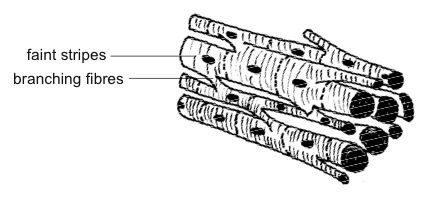
[edit | edit source]Cardiac muscle is found only in the walls of the heart where it produces the ‘heart beat’. Cardiac muscle cells are branched cylinders with central nuclei and faint stripes (see diagram 4.11). Each fibre contracts automatically but the heart beat as a whole is controlled by the pacemaker and the involuntary autonomic nervous system.

Nervous Tissues
[edit | edit source]Nervous tissue forms the nerves, spinal cord and brain. Nerve cells or neurons consist of a cell body and a long thread or axon that carries the nerve impulse. An insulating sheath of fatty material (myelin) usually surrounds the axon. Diagram 4.12 shows a typical motor neuron that sends messages to muscles to contract.
Vertebrate Bodies
[edit | edit source]We are so familiar with animals with backbones (i.e. vertebrates) that it seems rather unnecessary to point out that the body is divided into three sections. There is a well-defined head that contains the brain, the major sense organs and the mouth, a trunk that contains the other organs and a well-developed tail. Other features of vertebrates may be less apparent. For instance, vertebrates that live on the land have developed a flexible neck that is absent in fish where it would be in the way of the gills and interfere with streamlining. Mammals but not other vertebrates have a sheet of muscle called the diaphragm that divides the trunk into the chest region or thorax and the abdomen.
Body Cavities
[edit | edit source]
In contrast to many primitive animals, vertebrates have spaces or body cavities that contain the body organs. Most vertebrates have a single body cavity but in mammals the diaphragm divides the main cavity into a thoracic and an abdominal cavity. In the thoracic cavity the heart and lungs are surrounded by their own membranes so that cavities are created around the heart - the pericardial cavity, and around the lungs – the pleural cavity (see diagram 4.13).
Organs
[edit | edit source]
Just as the various parts of the cell work together to perform the cell’s functions and a large number of similar cells make up a tissue, so many different tissues can “cooperate” to form an organ that performs a particular function. For example, connective tissues, epithelial tissues, muscle tissue and nervous tissue combine to make the organ that we call the stomach. In turn the stomach combines with other organs like the intestines, liver and pancreas to form the digestive system (see diagram 4.14).
Generalised Plan Of The Mammalian Body
[edit | edit source]
At this point it would be a good idea to make yourself familiar with the major organs and their positions in the body of a mammal like the rabbit. Diagram 4.15 shows the main body organs.
Body Systems
[edit | edit source]Organs do not work in isolation but function in cooperation with other organs and body structures to bring about the MRS GREN functions necessary to keep an animal alive. For example the stomach can only work in conjunction with the mouth and esophagus (gullet). These provide it with the food it breaks down and digests. It then needs to pass the food on to the intestines etc. for further digestion and absorption. The organs involved with the taking of food into the body, the digestion and absorption of the food and elimination of waste products are collectively known as the digestive system.
The 11 body systems
[edit | edit source]- Skin
- The skin covering the body consists of two layers, the epidermis and dermis. Associated with these layers are hairs, feathers, claws, hoofs, glands and sense organs of the skin.
- Skeletal System
- This can be divided into the bones of the skeleton and the joints where the bones move over each other.
- Muscular System
- The muscles, in conjunction with the skeleton and joints, give the body the ability to move.
- Cardiovascular System
- This is also known as the circulatory system. It consists of the heart, the blood vessels and the blood. It transports substances around the body.
- Lymphatic System
- This system is responsible for collecting and “cleaning” the fluid that leaks out of the blood vessels. This fluid is then returned to the blood system. The lymphatic system also makes antibodies that protect the body from invasion by bacteria etc. It consists of lymphatic vessels, lymph nodes, the spleen and thymus glands.
- Respiratory System
- This is the system involved with bringing oxygen in the air into the body and getting rid of carbon dioxide, which is a waste product of processes that occur in the cell. It is made up of the trachea, bronchi, bronchioles, lungs, diaphragm, ribs and muscles that move the ribs in breathing.
- Digestive System
- This is also known as the gastrointestinal system, alimentary system or gut. It consists of the digestive tube and glands like the liver and pancreas that produce digestive secretions. It is concerned with breaking down the large molecules in foods into smaller ones that can be absorbed into the blood and lymph. Waste material is also eliminated by the digestive system.
- Urinary System
- This is also known as the renal system. It removes waste products from the blood and is made up of the kidneys, ureters and bladder.
- Reproductive System
- This is the system that keeps the species going by making new individuals. It is made up of the ovaries, uterus, vagina and fallopian tubes in the female and the testes with associated glands and ducts in the male.
- Nervous System
- This system coordinates the activities of the body and responses to the environment. It consists of the sense organs (eye, ear, semicircular canals, and organs of taste and smell), the nerves, brain and spinal cord.
- Endocrine System
- This is the system that produces chemical messengers or hormones. It consists of various endocrine glands (ductless glands) that include the pituitary, adrenal, thyroid and pineal glands as well as the testes and ovary.
Homeostasis
[edit | edit source]All the body systems, except the reproductive system, are involved with keeping the conditions inside the animal more or less stable. This is called homeostasis. These constant conditions are essential for the survival and proper functioning of the cells, tissues and organs of the body. The skin, for example, has an important role in keeping the temperature of the body constant. The kidneys keep the concentration of salts in the blood within limits and the islets of Langerhans in the pancreas maintain the correct level of glucose in the blood through the hormone insulin. As long as the various body processes remain within normal limits, the body functions properly and is healthy. Once homeostasis is disturbed disease or death may result. (See Chapters 12 and16 for more on homeostasis).
Directional Terms
[edit | edit source]
In the following chapters the systems of the body in the list above will be covered one by one. For each one the structure of the organs involved will be described and the way they function will be explained.
In order to describe structures in the body of an animal it is necessary to have a system for describing the position of parts of the body in relation to other parts. For example it may be necessary to describe the position of the liver in relation to the diaphragm, or the heart in relation to the lungs. Certainly if you work further with animals, in a veterinary clinic for example, it will be necessary to be able to accurately describe the position of an injury. The terms used for this are called directional terms.
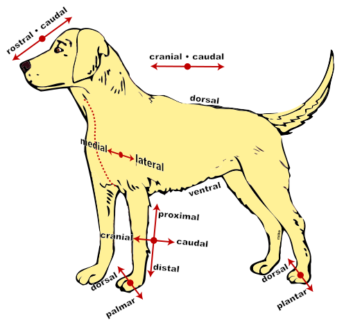
The most common directional terms are right and left. However, even these are not completely straightforward especially when looking at diagrams of animals. The convention is to show the left side of the animal or organ on the right side of the page. This is the view you would get looking down on an animal lying on its back during surgery or in a post-mortem. Sometimes it is useful to imagine ‘getting inside’ the animal (so to speak) to check which side is which. The other common and useful directional terms are listed below and shown in diagram 4.16.
| Term | Definition | Example |
|---|---|---|
| Dorsal | Nearer the back of the animal than | The backbone is dorsal to the belly |
| Ventral | Nearer the belly of the animal than | The breastbone is ventral to the heart |
| Cranial (or anterior) | Nearer to the skull than | The diaphragm is cranial to the stomach |
| Caudal (or posterior) | Nearer to the tail than | The ribs are caudal to the neck |
| Proximal | Closer to the body than (only used for structures on limbs) | The shoulder is proximal to the elbow |
| Distal | Further from the body than (only used for structures on limbs) | The ankle is distal to the knee |
| Medial | Nearer to the midline than | The bladder is medial to the hips |
| Lateral | Further from the midline than | The ribs are lateral to the lungs |
| Rostral | Towards the muzzle | There are more grey hairs in the rostral part of the head |
| Palmar | The "walking" surface of the front paw | There is a small cut on the left palmar surface |
| Plantar | The "walking" surface of the hind paw | The pads are on the plantar side of the foot |
Note that we don’t use the terms superior and inferior for animals. They are only used to describe the position of structures in the human body (and possibly apes) where the upright posture means some structures are above or superior to others.
In order to look at the structure of some of the parts or organs of the body it may be necessary to cut them open or even make thin slices of them that they can be examined under the microscope. The direction and position of slices or sections through an animal’s body have their own terminology.
If an animal or organ is sliced lengthwise this section is called a longitudinal or sagittal section. This is sometimes abbreviated to LS.
If the section is sliced crosswise it is called a transverse or cross section. This is sometimes abbreviated to TS or XS (see diagram 4.17).
Summary
[edit | edit source]- The characteristics of living organisms can be summarised by the words “MRS GREN.”
- There are 4 main types of tissue namely: epithelial, connective, muscle and nervous tissues.
- Epithelial tissues form the skin and line the gut, respiratory tract, bladder etc.
- Connective tissues form tendons, ligaments, adipose tissue, blood, cartilage and bone, and are found in the dermis of the skin.
- Muscular tissues contract and consist of 3 types: smooth, skeletal and cardiac.
- Vertebrate bodies have a head, trunk and tail. Body organs are located in body cavities. 11 body systems perform essential body functions most of which maintain a stable environment or homeostasis within the animal.
- Directional terms describe the location of parts of the body in relation to other parts.
Worksheets
[edit | edit source]Students often find it hard learning how to use directional terms correctly. There are two worksheets to help you with these and another on tissues.
Test Yourself
[edit | edit source]1. Living organisms can be distinguished from non-living matter because they usually move and grow. Name 5 other functions of living organisms:
- 1.
- 2.
- 3.
- 4.
- 5.
2. What tissue types would you find...
- a) lining the intestine:
- b) covering the body:
- c) moving bones:
- d) flowing through blood vessels:
- e) linking the eye to the brain:
- f) lining the bladder:
3. Name the body cavity in which the following organs are found:
- a) heart:
- b) bladder:
- c) stomach:
- d) lungs:
4. Name the body system that...
- a) includes the bones and joints:
- b) includes the ovaries and testes:
- c) produces hormones:
- d) includes the heart, blood vessels and blood:
5. What is homeostasis?
6. Circle which is correct:
- a) The head is cranial | caudal to the neck
- b) The heart is medial | lateral to the ribs
- c) The elbow is proximal | distal to the fingers
- d) The spine is dorsal | ventral to the heart
7. Indicate whether or not these statements are true.
- a) The stomach is cranial to the diaphragm - true | false
- b) The heart lies in the pelvic cavity - true | false
- c) The spleen is roughly the same size as the stomach and lies near it - true | false
- d) The small intestine is proximal to the kidneys - true | false
- e) The bladder is medial to the hips - true | false
- f) The liver is cranial to the heart - true | false
Websites
[edit | edit source]- Animal organ systems and homeostasis
Overview of the different organ systems (in humans) and their functions in maintaining homeostasis in the body.
http://www.emc.maricopa.edu/faculty/farabee/biobk/BioBookANIMORGSYS.html
- Wikipedia
Directional terms for animals. A little more detail than required but still great.
http://en.wikipedia.org/wiki/Anatomical_terms_of_location
