Anatomy and Physiology of Animals/Cardiovascular System/The Heart
Objectives | The Heart
[edit | edit source]After completing this section, you should know:
- where the heart is located in the body
- the structure of the heart
- the structure and function of the heart valves and their role in producing the heart sounds
- the stages of the heart beat and the route the blood takes through the heart
- that the coronary arteries supply the heart muscle
The Heart
[edit | edit source]
The heart is the pump that pushes the blood around the body in the blood vessels of the circulatory system. In fish the blood only passes through the heart once on its way to the gills and then round the rest of the body. However, in mammals and birds that have lungs, the blood passes through the heart twice: once on its way to the lungs where it picks up oxygen and then through the heart again to be pumped all over the body. The heart is therefore two separate pumps, side by side (see diagram 8.6).
The heart is situated in the thorax between the lungs and is protected by the rib cage. In some animals it is displaced slightly to the left-hand side. A tough membrane called the pericardium covers it. There is a narrow space between the pericardium and the heart that is filled with a liquid that acts as a lubricant.
The heart of mammals is a hollow bag made of cardiac muscle (see chapter 4). The cavity inside the heart is divided into 4 chambers. The chambers on the right side are completely separate from the chambers on the left side. The two upper chambers are thin walled, and are called the atria (or auricles). The two lower chambers are thick walled and are called the ventricles (see diagrams 8.7 and 8.8).
 |
The Heart
[edit | edit source]Blood flows through the heart in a one way system. The right atrium receives deoxygenated blood from the body via the largest vein in the body called the vena cava. The contraction of the atrium pumps the blood into the right ventricle and then into the lungs via the pulmonary artery. The blood is oxygenated in the lungs and then returns to the heart and enters the left atrium via the pulmonary vein. The contraction of the left atrium pumps the blood into the left ventricle, which then pumps it to the body via the aorta (see diagrams 8.7 and 8.8). The wall of the left ventricle is usually much thicker than that of the right ventricle because it has to pump the blood to the end of the digits and tip of the tail while the right ventricle only has to pump the blood to the nearby lungs.
Valves
[edit | edit source]Valves are flaps of tissue that stop blood flowing backwards and so control the direction of blood flow in the heart. There are two kinds of valves in the heart. The first kind is the massive valves between the atria and the ventricles, the atrio-ventricular valves, (AV valves) that prevent blood in the ventricles from flowing back into the atria. The flaps of these valves are attached to the walls of the ventricles by tendons. These make them look somewhat like parachutes (see diagram 8.9).
Diagram 8.9 - The atrio-ventricular or parachute valves
The second kind of valve is pocket shaped flaps of tissue called the semilunar (half moon) valves (see diagram 8). They are called the pulmonary and aortic valves and found at the back of the pulmonary artery and aorta respectively.
The Heartbeat
[edit | edit source]The heartbeat consists of alternating contractions and relaxations of the heart. If you listen to the heart with a stethoscope, you hear the sounds often described as “lub-dub”.
There are four stages to each heartbeat:
- Each atrium relaxes so that blood can enter. Blood flows from the body via the vena cava into the right atrium. At the same time, blood flows from the lungs via the pulmonary vein into the left atrium (see diagram 8.10a).
- The atrioventricular valves open and both ventricles relax. The atria contracts and blood flows from the right atrium into the right ventricle and from the left atrium into the left ventricle (see diagram 8.10b).
- The ventricles contract and the atrioventricular valves snap shut to stop blood flowing back into the atria. This is the first sound (“lub”) of the heartbeat that can be heard with a stethoscope (see diagram 8.10c).
- The semi-lunar valves open and blood is pumped out of the right ventricle to the lungs. At the same time, blood is pumped out of the left ventricle into the aorta and so to the rest of the body. When the ventricles stop contracting, the semi-lunar valves snap shut to stop blood flowing backward.
This is the second sound (“dub”) of the heartbeat. Blood flows into the atria again as they relax and the cycle is repeated.
When a valve is damaged and fails to close completely, some blood may flow backward after each heartbeat. A trained veterinarian hears this with a stethoscope as a “heart murmur”.
The period of the heartbeat when the ventricles are contracting and sending a wave of blood down the pulmonary artery and aorta is called systole. The period when the ventricles are relaxing is called diastole.
Cardiac Muscle
[edit | edit source]The walls of the heart consist of cardiac muscle, a special kind of muscle only found in the heart. The cells of cardiac muscle form a branching network of separating and rejoining fibres which allows nerve impulses to travel through the tissue (see Chapter 7). Heart muscle needs lots of energy to function so it is well supplied with mitochondria and requires a good supply of oxygen. This is provided by the coronary arteries (see below).
Diagram 8.10 a) First stage of heartbeat b) Second stage of heartbeat c) Third stage of heartbeat
Control Of The Heartbeat
[edit | edit source]The cardiac muscle of the walls of the heart contracts of its own accord. This can be demonstrated by the rather macabre experiment in which a small portion of heart muscle is removed and placed in a solution that is similar to blood. The tissue will continue to contract and relax for a time. In the normal functioning heart the pacemaker acts rather like the conductor of an orchestra and superimposes a unified beat upon the heart as a whole. The pacemaker is situated in the wall of the right atrium. The rate at which the heart beats is modified by a part of the brain called the medulla oblongata (see Chapter 14) and by the hormone adrenalin (see Chapter 16) which speeds up the heartbeat.
The Coronary Vessels
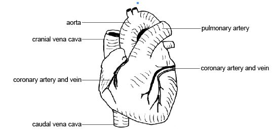
[edit | edit source]Although oxygenated blood passes through some of the chambers of the heart it can not supply the muscle of the heart walls with the oxygen and nutrients it needs. Special arteries called the coronary arteries do this. These two arteries arise from the aorta and branch through the heart to deliver oxygen and nutrients to the cardiac muscles and collect carbon dioxide and wastes. Coronary veins return the blood to right hand side of the heart. Some of these vessels can be seen on the outside surface of the heart (see diagram 8.11). Sometimes fatty deposits on the inside wall of the coronary artery block the blood flow to the heart muscle. If the obstruction is severe enough to damage the heart muscle due to inadequate blood supply a “heart attack” can result.
Diagram 8.11 - The heart showing coronary vessels
Summary
[edit | edit source]- The heart is situated in the thorax between the lungs
- The heart is a hollow bag made of cardiac muscle. It is divided into four chambers (right and left atria and right and left ventricles).
- Valves stop blood flowing backwards. The right and left atrio-ventricular valves prevent blood in the ventricles from flowing back into the atria. The semilunar valves at the entrance of the pulmonary artery and aorta prevent blood flowing back into the ventricles. The closing of the valves produces the heart sounds heard with a stethoscope.
- There are 4 stages to the heart beat. 1. blood flows into the right and left atria. 2. The atria contract and blood flows into the ventricles. 3. The ventricles contract and the closing of the atrio-ventricular valves produces the first heart sound. 4. Blood flows to the lungs and body and when the ventricles stop contracting the closing of the semilunar valves produces the second heart sound.
- The coronary arteries supply the heart muscle with oxygenated blood.
Worksheet
[edit | edit source]Use the Heart Worksheet to help you learn the different parts of the heart, the role of the heart valves and how the heart beat pushes the blood through the heart.
Test Yourself
[edit | edit source]- Via what vessel does blood enter the heart from the body?
- Blood passing through the right atrioventricular valve passes into which chamber of the heart?
- What is the function of the pulmonary valve?
- Does the pulmonary artery carry deoxygenated or oxygenated blood?
- The walls of the heart are made of what tissue?
- Does the aorta carry deoxygenated or oxygenated blood?
Websites on the Heart
[edit | edit source]- http://www.guidant.com/condition/heart/heart_bloodflow.shtml Blood flow through heart
An animation of blood flow through the heart with step-by-step annotated breakdown. Also an animation putting heartbeat, valve operation and blood flow all together.
Great animation showing parts of heart plus constituents of blood including RBCs, white cells, platelets and plasma. Even shows how to make a blood smear and identify the white cells on it as well as make and read a haematocrit. Some parts are a little too advanced.
- http://web.archive.org/web/20040523224602/http://www.bishopstopford.com/faculties/science/arthur/Heart%20drag&drop.swf Heart animation
Drag and drop animation of the human heart. Great for revision but note the terms bicuspid and tricuspid valves are used. These are equivalent to the left and right atrio-ventricular valves in animals.
A good animation of the heartbeat showing the valves opening and closing with appropriate heart sounds. Plus a good diagram showing the difference between the atrio-ventricular and semilunar valves.
- http://www.nucleusinc.com/animation2.php Human heart animation
A great animation where you use the mouse to point to parts of the heart and a voice over tells you what part it is. Just note this is of the human heart so the terms superior and inferior vena cava are used instead of caudal and cranial as for an animal.
- http://en.wikipedia.org/wiki/Heart The heart
Again Wikipedia is a wonderful resource, although remember, most of the material is on the human system.


