Cell Biology/History
Some History of the development of understanding of the Cell
[edit | edit source]The origin of the idea that living organisms are made of cells is often traced back to observations of thin slices of cork. In 1665 the book Micrographia: Some physiological descriptions of minute bodies made by magnifying glasses was published by Robert Hooke. He wrote:
- . . . I could exceedingly plainly perceive it to be all perforated and porous, much like a Honey-comb, but that the pores of it were not regular. . . . these pores, or cells, . . . were indeed the first microscopical pores I ever saw, and perhaps, that were ever seen, for I had not met with any Writer or Person, that had made any mention of them before this. . .
We now know that the "cells" Hooke observed were an indication of the cellular structure of multi-cellular organisms. During the 1670s, Antony van Leeuwenhoek used microscopes to observe sperm, red blood cells, and protozoa. While many cells are about 10 microns in diameter, some protozoa are visible to the naked eye, reaching over 1 millimeter in length. Leewenhoek is the inventor of the microscope. Thus, while it is true that the small size of most cells made it difficult to develop the theory that all living organisms are composed of cells, it was also difficult to recognize that living cells have certain functional components such as the nucleus and a surface membrane that allow cells to exist as the basic functional components of all living organisms. In 1833 Robert Brown published a report describing microscopic observations of plant cells in which he used first used the term "cell nucleus":
- In the compressed cells of the epidermis the nucleus is in a corresponding degree flattened; but in the internal tissue it is often nearly spherical, more or less firmly adhering to one of the walls, and projecting into the cavity of the cell.
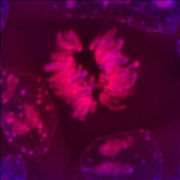
Such observations of the microscopic cellular components of cells helped make it possible for Schleiden and Schwann to propose a cell theory specifying that nucleated cells are key structural and functional units in plants and animals (1832-1838). However, they did not understand cell reproduction. About this time microscopists such as the Belgian botanist Barthelemy C. Dumortier observed and reported the binary fission of cells. By 1879 the zoologist Walther Flemming was using chemical staining of "fixed" cells to allow clear visualization of chromosomes during cell division.
During the 1890s, Ernest Overton, developed a theory of lipid membrane structure and function, based largely on the osmotic properties of cells. Visualization of lipid bilayer membranes at the surface of cells had to wait until the development of electron microscopy.
<< | Size of cells


