Learning anatomy/Table version
This will expand every day.
Learning anatomy
Learning anatomy is not completed. The learning anatomy crew is where you get to see all the authors. This is the fastest way to learn anatomy, even faster than kenhub plus you don't need to pay a single penny for any part of the book! Interested in the brain? Then check out the human brain book. Learning anatomy
A journey under the skin
Versions of learning anatomy There is a Table version of learning anatomy. ________________________________________________________________________________________________________________
Table of contents[edit | edit source]Gastrointestinal anatomyHepato-Pancreato-Biliary anatomyCardiovascular anatomyKidney UrinationMusculoskeletal anatomyEar, Nose and ThroatSkinLungOtherIf you finished the book go to the Test area to check your knowledge. External links[edit | edit source]
| |||
Esophagus
Anatomy[edit | edit source]The esophagus is a tube connecting the throat to the stomach. The parts of the esophagus are the upper esophagus, middle esophagus, and lower esophagus. The esophagus also passes through a hole in the diaphragm called the esophageal hiatus. Function[edit | edit source]The function of the esophagus is to move the swallowed food from the throat into the stomach. The lower esophageal sphincter or cardiac sphincter is between the lower esophagus and cardiac region of the stomach. Peristalsis happens throughout the gastrointestinal tract. | |||
Stomach
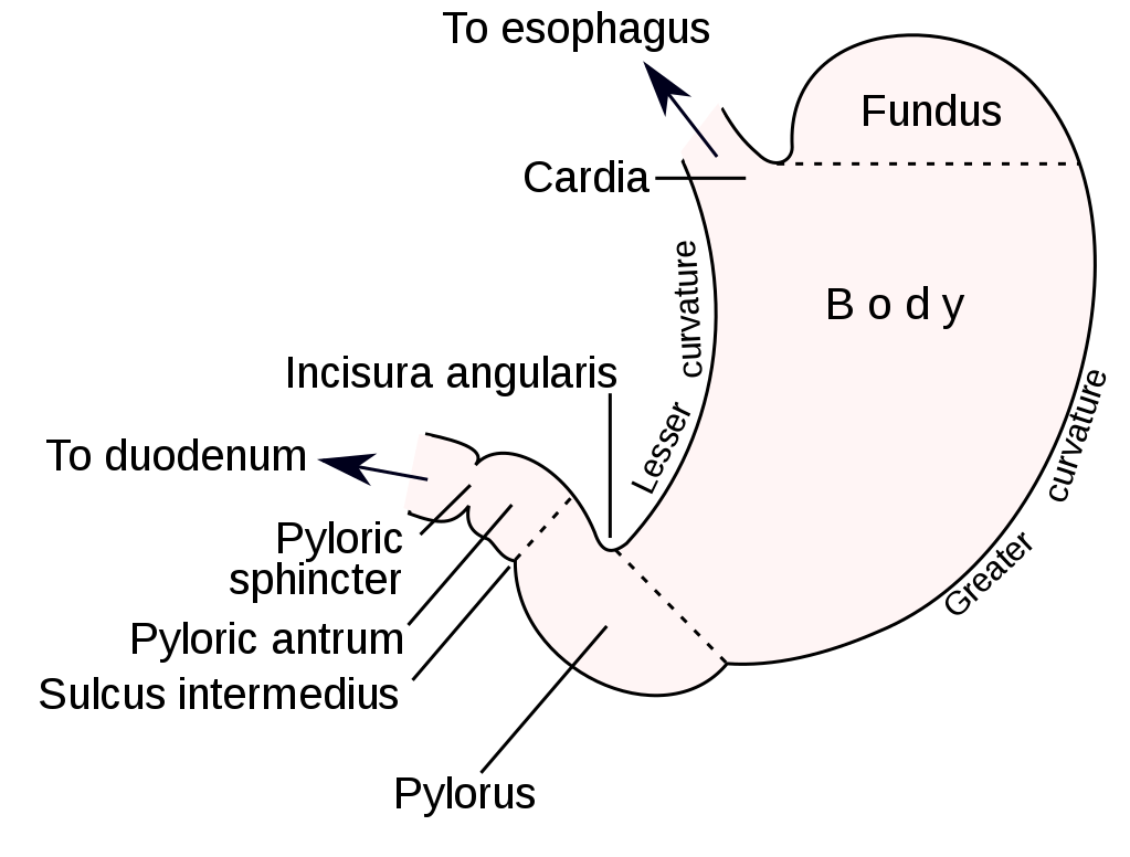
Anatomy[edit | edit source]The stomach is a J-shaped ”bag” that connects the esophagus to the small bowel. The parts of the stomach are the cardiac region, gastric fundus, gastric body, and pyloric region. The pyloric region is made up of the pyloric antrum, and the pyloric canal.
Function[edit | edit source]The first sphincter of the stomach is the cardiac sphincter or lower esophageal sphincter which guards the entrance to the stomach. If the sphincter weakens, stomach contents can damage the lining of the esophagus and cause acid reflux so, please take care of your lower esophageal sphincter. The stomach has folds called rugae which look wrinkled when no food is in the stomach. The stomach stores food. If the food is poisonous, it triggers the vomiting reflex. In this case, the body protects itself from the acid. But if it’s not poisonous, the food continues to go. The stomach will make acid, digestive enzymes, and mucin. Hydrochloric acid breaks down all foods into chyme. Amylase breaks down starch. Pepsinogen is the inactive form of pepsin. When pepsinogen reaches the acidic ph of the stomach, it turns into pepsin which digests proteins. Gastric lipase breaks down lipids. Hydrochloric acid in the stomach kills germs. Gastrin and somatostatin are hormones. Gastrin inhibits acid secretion. Somatostatin does a lot of things. | |||
Small bowel Stomach - Large bowel Template:Toc Anatomy[edit | edit source]The small bowel or small intestine connects the stomach to the large intestine. The parts of the small intestine are the duodenum, jejunum, and ileum. Function[edit | edit source]The small intestine absorbs most of the nutrients from the chyme. The pyloric sphincter lets only a tiny bit of chyme go into the small intestine at a time. The small finger-like projections are called villi. Villi help in the absorption. The jejunum absorbs sugars, amino acids, and fatty acids. The ileum absorbs bile acids, fluid, and vitamin B12. | |||
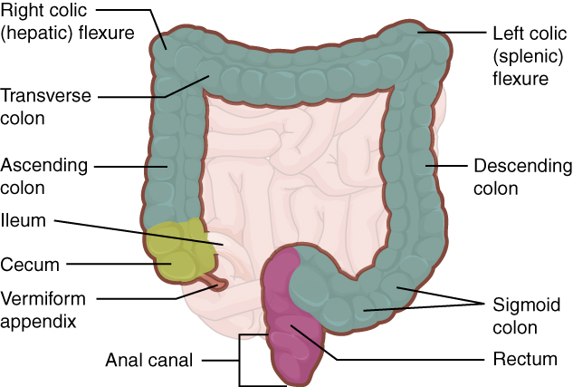
Large bowel Anatomy[edit | edit source]The large bowel or large intestine is made up of the vermiform appendix, the cecum, and the colon. The colon is made up of the ascending colon, transverse colon, descending colon, and sigmoid colon. The large intestine connects the small intestine to the rectum.
Function[edit | edit source]The cecum absorbs fluids and salts. The vermiform appendix is a storage for the gut bacteria that help with digestion. Gut bacteria is an example of a good bacteria. The colon absorbs water and electrolytes. | |||
Rectum and anus Anatomy[edit | edit source]The rectum and anus connect the large intestine to the external body. There is also an anal canal that goes between them. Defecation[edit | edit source]The function of the rectum and anus is defecation. In this section, you will learn about the defecation reflex. The rectum stores the feces that the colon made. The nerve signals then tell the brain that you need to go into the bathroom. Then the feces go into the anus. There are 2 sphincters. The first is the internal sphincter and we can't control it. The last one is the external sphincter and we control it. Babys can't control their external sphincter so they do it in their diapers. | |||
Liver Accessory organs _______________________________________________________________________
Anatomy[edit | edit source]The liver is made up of the left lobe, right lobe, caudate lobe, and quadrate lobe. The ligaments are the falciform ligament and the coronary ligament. They are not real ligaments. Instead, they are a piece of peritoneal membrane holding the liver. The lobes of the liver are divided into 8 segments.
Function[edit | edit source]The liver has many functions and is a critical part of the digestive system. Did you know that the liver has more than 500 functions and can do 200 functions at the same time!? The liver makes bile, stores nutrients and performs metabolism. Bile is made by hepatocytes which are also called liver cells. | |||
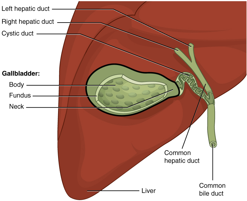
Biliary tract Accessory organs _______________________________________________________________________ Anatomy[edit | edit source]The biliary tract is made up of the liver, gallbladder, and bile ducts.
Function[edit | edit source]The hepatocytes make bile. After that, the bile goes into the bile canaliculi and then the bile goes into the interlobular ducts and then the bile goes into the intrahepatic ducts and then the bile moves into the right and left hepatic ducts and then the bile moves into the common hepatic duct and then the bile goes into the cystic duct and then the bile goes into the gallbladder. The gallbladder stores bile until needed. When needed, cholecystokinin triggers the bile to go out of the gallbladder and then into the common bile duct. Then the bile moves into the duodenum where the bile emulsifies fat. | |||
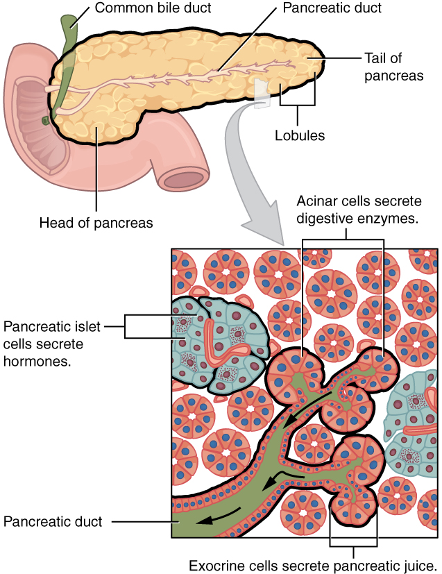
Pancreas exocrine Accessory organs _______________________________________________________________________ Biliary tract - Pancreas endocrine Anatomy[edit | edit source]Function[edit | edit source]Acinar cells make digestive enzymes and bicarbonate. Bicarbonate neutralizes the hydrochloric acid from the stomach. Amylase breaks down starch. Lipase breaks down lipids. Trypsin breaks down proteins. Chymotrypsinogen is activated by trypsin to make chymotrypsin. Chymotrypsin breaks proteins in the chyme. | |||
Pancreas endocrine Accessory organs _______________________________________________________________________ Anatomy[edit | edit source]Endocrine cells in the pancreas.
Function[edit | edit source]Beta cells make insulin. Alpha cells make glucagon. Insulin is like a key for opening up the cells so that glucagon can get in. | |||
| Example | |||
| Example | |||
| Example | |||
| Example | |||
| Example | |||
| Example | |||
| Example | |||
| Example | |||
| Example | |||
| Example | |||
| Example | |||
| Example | |||
| Example | |||
| Example | |||
| Example | |||
| Example | |||
| Example | |||
| Example | |||
| Example | |||
| Example | |||
| Example | |||
| Example | |||
| Example | |||
| Example | |||
| Example | |||
| Example | |||
| Example | |||
| Example | |||
| Example |
This is the Table version of Learning anatomy.


