Medical Physiology/Basic Biochemistry/Nucleic Acids
Introduction & Overview
[edit | edit source]Cells are divided into two broad categories, Prokaryocytes and Eukaryocytes. Basically, Prokaryocytes have no nucleus, and all bacteria fall into this category; eukaryocytes have a nucleus, and animal and Plant cells fall into this category. A characteristic of both cells is that they contain all the information necessary to build a complete organism. This information is carried in the cell's DNA. In prokaryocytes this exists as a ring of DNA which is found in the cell cytoplasm, in the Animal the DNA is found in the nucleus and is organized into chromosomes. Both types of DNA are organised into genes. A gene is the sequence of Nucleic acids necessary to produce a protein. This section looks at the Chemistry of DNA and RNA.
We will look at the chemical structure of Nucleic Acids and related compounds. We will only touch briefly on protein synthesis and replication. we will look at this in much more detail in the Section on Cell Biology particularly in the sections on Gene Expression and Cell Reproduction.
DNA Structure
[edit | edit source]A DNA molecule consists of two polymers of nucleic acids: the polymers consist of strands of nucleic acids joined together. There are thus two strands of DNA which run in opposite directions - they are said to be anti-parallel - and these strands are joined to each other by hydrogen bonds. (See below for the details). The weakness of these bonds allows the strands to be easily separated for reproduction and for the manufacture of RNA. There are four nucleic acids found in DNA, and these are grouped together in triplets called codons. Each codon is specific for a particular amino acid.
DNA Function
[edit | edit source]DNA controls the whole functioning of the cell. Although each cell contains an entire copy of the body's DNA, only some of the DNA is expressed in any given cell. The expression is both temporal and spatial and will also vary according to the needs of the body. It is carried out by way of manufacturing proteins, both structural proteins and enzymes.
                                                 /---->Cell Enzymes------>\
Gene(DNA) --->RNA Formation--->Protein Formation                            ---> Cell Function
                                                 \---->Cell Structure ---->/
The importance of DNA lies in its ability to control the formation of proteins via the genetic code. It does this by way of codons which as already mentioned is a triplet of nucleic acids. When a signal arrives to create a particular protein, the relevant section of DNA is identified, and an enzyme called RNA polymerase causes the two strands break apart. DNA of one of the strands, called the transcription strand serves as a template for the manufacture of messenger RNA (mRNA), a process called transcription. This strand of RNA contains a mirror image of all the codons each of which represents an amino acid.
After further processing the mRNA passes out of the nucleus through membrane pores and attaches itself to a ribosome. A second form of RNA called transfer RNA (tRNA) then ferries amino acids to the mRNA which assembles them into a poly peptide of the sequence dictated initially in the DNA of the gene. This process whereby a protein is manufactured from an mRNA strand is called Translation. Proteins are usually then taken inside the endoplasmic reticulum for further processing.
Protein Synthesis
[edit | edit source]The following illustrations show in simplified form how protein synthesis is carried out. More details are given in the section on Gene expression in the Cell Biology section.
A mRNA strand is transcribed from the DNA template; it is further modified in the nucleus; then it leaves the nucleus and attaches to a ribosome; then the peptide is translated from this mRNA strand. This peptide can be further modified in the endoplasmic reticulum.
RNA Transcription
[edit | edit source]This illustration shows a summary of the Transcription Process;
RNA Translation
[edit | edit source]This illustration shows a summary of the Translation Process
This illustration shows tRNA ferrying amino acids to the mRNA template

This is a very simple overview and will be look at in more detail when we consider Gene Expression in a later chapter.
Chemistry and Structure
[edit | edit source]Basic Structure
[edit | edit source]Nucleotides are three-part molecules that link together to form DNA (Deoxyribose Nucleic Acid) or RNA (Ribose Nucleic Acid) polymers. Each nucleotide is a three-part molecule consisting of one or more phosphate groups; a five-carbon sugar, and a carbon nitrogen ring called a base. Here is a diagram of the nucleic acid Guanine:
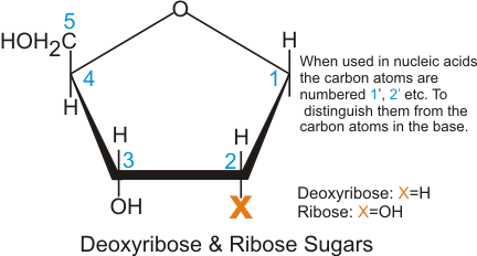
Note the numbering of the carbon atoms on the ribose ring. These nucleotides can be linked by their phosphate groups to form either an RNA or a DNA polymer.
Nucleotide Bases
[edit | edit source]

There are five Nucleotide bases, based on either a pyrimidine or a purine model. The purine derivatives have two carbon-nitrogen rings and form the bases adenine and guanine. The pyrimidine derivatives have one carbon-nitrogen ring and form the bases cytosine, thymine and uracil. The Thymine base appears only in DNA molecules, the Uracil base in RNA molecules.
Pentose sugars
[edit | edit source]The ribose sugar is used in RNA molecules, the Deoxyribose sugar in DNA molecules
DNA and RNA Structure
[edit | edit source]The DNA Molecule
[edit | edit source]The DNA molecule arranges itself in the famous double helix, first postulated by Watson and Crick in 1953:
DNA Structure
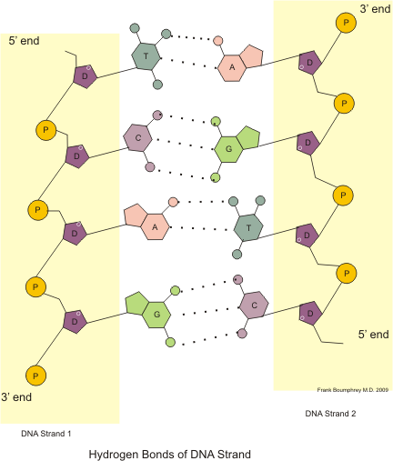
[edit | edit source]DNA consists of two chains of DNA polymers linked by Hydrogen bonds. Each chain has either a terminal phosphate or a terminal Ribose group. The end with phosphate group is known as the 3 prime - 3' end, that with the pentose group as the 5 prime - 5' end. This distinction is important when we have a look at DNA and RNA replication.
The chains run in opposite directions, one from 3' to 5', the other from 5' to 3'.
The following illustration shows how the individual Nucleic Acids link together in a single strand of DNA:
In the chain an Adenine base always matches with a Thymine base, and a Guanine base with a Cytosine base.
The following Illustration shows a DNA Strand combined with a reverse strand. They are held together by Hydrogen Bonds (the dotted lines) between complementary nucleic acids:
- Guanine always matches with Cytosine.
- Adenine always matches with Thymine.
Remember this match with the mnemonic "The Audience Goes Crazy"
- Thymine and Adenine link with two hydrogen bonds.
- Guanine and Cytosine link with three hydrogen bonds.
Here is another shorthand method of depicting DNA:
RNA Structure
[edit | edit source]RNA molecules are formed using the Ribose sugar. They do not form into helixes. Another difference is that they do not have Thymine bases, they use Uracil in its place. They use a DNA template and are formed from the 5' to the 3' end. Here is a structural look at the DNA helix and an RNA strand:
The Nucleic Acids are added one at a time in the tri-phosphate form. First the nucleic acid is matched with its opposite number, then an ester bond is formed between the hydroxyl group at the 3' position and the first phosphate group, cleaving off a pyrophosphate. The energy released by this cleavage powers the reaction.
Again note that the RNA strand is started at the 5' end and works along the DNA template from the 3' end to the 5' end.
There are numerous kinds of RNA
- mRNA which we have just had a look at
- Primer RNA which is important in the process of DNA replication
- Transfer RNA (tRNA) which ferries amino acids to the mRNA strand during the translation process.
- Ribosomal RNA, which forms a large part of the ribosomes
DNA Replication
[edit | edit source]Here we give a simplified overview of DNA replication, emphasizing the chemistry. We will give a much more detailed view when we consider cell reproduction in the Cell Biology section.
When DNA is replicated, the whole chromosome is replicated. As this contains up to 9 billion base pairs, if the replication started at one end and then proceeded to the other it would take an inordinate amount of time. Replication therefore proceeds at numerous locations at the same time. Bubbles are formed, and replication proceeds at both ends of the bubble at replication forks.
Nucleoside triphosphates are added on at the 3' end of the replication chain. One of the DNA Polymerases enzymes (δDNA Poly) facilitates the reaction.
- First the Base pairs with its opposite number in the template chain, forming a hydrogen bond.
- Next the first phosphate bond of the nucleoside triphosphate joins with the hydroxyl group at the 3' carbon.
The energy obtained from cleaving off the last two phosphates powers the reaction.
Both during and after the formation of the replication chain, various enzymes check for accuracy of transcription, and carry out the necessary repairs. This, as well as the numerous activities at the replication fork, and telomere activities at the end of the chain will be looked at in further detail in the section on cell replication.


