Medical Physiology/Gastrointestinal Physiology/Digestion & Absorption
Digestion and Absorption
[edit | edit source]Overview
[edit | edit source]Digestion of food breaks the large molecules into smaller molecules suitable for absorbing in the small intestine. This takes place either both in the lumen of the canal in the chyme and at the epithelial junction of the cells of the small intestine.
The surface area for absorption is greatly increased by the christal folds and the villi of the small intestine (see anatomy), and the microvilli of the epithelial cells them selves, which form the brush border.
As was mentioned in the secretion section, if digestive secretions of the small intestine are collected free of cell debris then no enzymes are seen. We now know that the small intestine enzymes are anchored in the apical (lumen) margin of the epithelial cell (enterocyte), in the brush border. This prevents them from being washed down stream with the chyme
The Enterocyte is specialized for absorption of food stuffs, it is divided into an apical or lumenal surface where the final digestion and absorption takes place, and a basal/lateral surface surface where the products of digestion are passed into the interstitial fluid. The transport mechanisms at these two surfaces are quite different. The apical surface is characterized by numerous microvilli which greatly increase the surface area available for absorption. Adjacent to this is an unstirred layer of mucous that the products of digestion must penetrate before being absorbed.
In healthy intestines, the junctions between the cells are very tight, and no leakage takes place. At the walls at the base of the cells there is a space continuous with the interstitial space.
The enzymes of the small intestine consist of several peptidases; several enzymes for splitting disaccharides into monosaccharides, and a lipase. These enzymes operate while the substrates are being absorbed through the epithelium.
In this chapter we will look at the digestive mechanisms of food stuffs, and then at the mechanics of how the products are absorbed into the body.
Carbohydrate Digestion
[edit | edit source]Carbohydrates are digested by salivary and pancreatic enzymes (α-amylases) and by numerous oligosaccharidases of the small intestine wall. The following table shows the enzymes that operate on starch:
| Source | Enzyme | Activated by | Substrate | Functions and/or Products |
|---|---|---|---|---|
| Saliva | Salivary α-amylase (Ptyalin) | Cl- | Starch | Hydrolyses non-terminal α1:4 linkages to produce dextrins, maltotriose, and maltose |
| Pancreas | Pancreatic α-amylase (Ptyalin) | Cl- | Starch | Hydrolyses non-terminal α1:4 linkages to produce dextrins, maltotriose, and maltose |
| Small Intestine mucosa | ... | Glucose | ||
| Maltase | ... | Maltose, maltotriose, α-dextrins | Glucose | |
| Lactase | ... | Lactose | Glucose and Galactose | |
| Sucrase | ... | Sucrose, maltose, maltotriose | Glucose | |
| α-Dextrinase | ... | α-dextrins, maltose, maltotriose | Glucose | |
| Trehalase | ... | Trehalose | Glucose |
The amylase of the salivary gland (a.k.a.Ptyalin) is inactivated by stomach acid, but when food enters the stomach it is first stored in the fundus of the stomach and not mixed with acid, so ptyalin can carry on acting until mixing with stomach acid takes place, which can be sometime. By some estimates up to a third of the carbohydrates are reduced to polysaccharides by the time the chyme leaves the stomach. The other two thirds are digested by pancreatic amylase.
Carbohydrates are taken in in mainly the form of plant carbohydrate (amylose) and animal carbohydrate (glycogen) together with some sugars, mainly disaccharides . In the western diet about 80% is in the form of amylose. Amylose is not highly branched and consists mainly of long chains of glucose linked by α1:4 linkages. Cellulose, the most abundant starch in nature, is formed of β1:4 linkages and cannot be digested in humans, although bacterial action in the colon does breakdown a minute amount.
Glycogen is a multi-branched starch with linkages at the 1:4 and 1:6 position.
This gives very large granules of multi-branched starch.
Both the parotid and pancreatic amylases hydrolyse the 1:4 link, but not the terminal 1:4 links or the 1:6 links. This breaks amylose down into mainly disaccharides, and glycogen with its 1:6 linkages into polysaccharides.
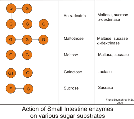
The net result of these actions are numerous disaccharides and polysaccharides. These are broken down into monosaccharides by the enzymes attached to the enterocycytes of the small intestine, as illustrated below:
Carbohydrate Absorption
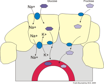
[edit | edit source]Glucose is transported from the lumen into the cell by a sodium linked co-transporter or symport (SGLT) and is thus highly dependent on the concentration of sodium in the lumen. Galactose uses the same mechanism. (see section on cell membrane transport for further details)
Fructose uses a different mechanism, a facilitated diffusion carrier and is sodium independent.
Glucose is carried across the baso-lateral membrane by a facilitated diffusion carrier.
The sugars are absorbed in the capillaries and carried to the liver.
Protein Digestion
[edit | edit source]Proteins and polypeptides are digested by hydrolysis of the C-N bond.
The proteolytic enzymes are all secreted in an inactive form, to prevent auto-digestion, and are activated in the lumen of the gut: by HCl in the case of the stomach pepsinogen; by enteropeptidase and trypsin in the case of the pancreatic enzymes. This was discussed in the section on secretions. Final digestion takes place by small intestine enzymes embedded in the brush border of the small intestine.
The enzymes are divided into endo- and exo-peptidases. The endopeptidases cleave the polypeptide at the interior peptide bonds, the exopeptidases cleave the terminal amino acid. Exopeptidases are further subclassified into aminopeptidases - which cleave off the terminal amino acid at the amine end of the chain, and carboxypeptidases which cleave off the terminal amino acid at the carboxyl end of the chain. This is illustrated graphically here:
Stomach pepsin cleaves interior bonds of the amino acids, and is particularly important for its ability to digest collagen. This is a major constituent of connective tissue of meat. In the absence of stomach pepsin, digestion in the small intestine proceeds with difficulty. Stomach pepsin digests about 20% of the proteins, the rest is digested by pancreatic and small intestine enzymes.
| Source | Enzyme | Activated by | Substrate | Functions and/or Products |
|---|---|---|---|---|
| Stomach | Pepsins (pepsinogens) | HCl | Proteins and Polypeptides | Cleaves interior peptide bonds of aromatic amino acids^^ |
| Pancreas | Trypsin (trypsinogen) | Enteropeptidase | Proteins and Polypeptides | Cleaves peptide bonds on carboxyl side of basic amino acids^ (argenine or Lysine) |
| Chymotrypsin (chymotrypsinogen) | Trypsin | Proteins and Polypeptides | Cleaves peptide bonds on carboxyl side of aromatic amino acids^^ | |
| Elastase (proelastase) | Trypsin | Elastin and other Proteins | Cleaves peptide bonds on carboxyl side of aliphatic amino acids^^^ | |
| Carboxypeptidase A (procarboxypeptidase A) | Trypsin | Proteins and Polypeptides | Cleaves peptide bonds on carboxyl terminal amino acids with aliphatic or aromatic side chains | |
| Carboxypeptidase B (procarboxypeptidase B) | Trypsin | Proteins and Polypeptides | Cleaves peptide bonds on carboxyl terminal amino acids with basic side chains | |
| Small Intestine mucosa | Enteropeptidase | ... | Trypsinogen | Trypsin |
| Aminopeptidases | ... | Polypeptides | Cleaves amino terminal amino acid from peptide | |
| Carboxypeptidases | ... | Polypeptides | Cleaves carboxyl terminal amino acid from peptide | |
| Endopeptidases | ... | Polypeptides | Cleaves mid portions of peptide | |
| Dipeptidases | ... | Dipeptides | ----> two amino acids |
^ Aromatic amino acids are amino acids which include an aromatic ring. They include phenylalanine, histidine, tryptophan, and tyrosine.
^^ Basic amino acids are polar and positively charged at pH values below their pKa's, and are very hydrophilic. They include histadine, lysine and argenine.
^^^ Aliphatic implies that the protein side chain contains only carbon or hydrogen atoms.
Protein Absorption
[edit | edit source]Several different transport systems transport amino acids into the enterocyte, each dealing with different groups of amino acids. Most of these are sodium dependent co-transporters. Di-peptides and tri-peptites are transported by a H+ ion dependent co-transporter. In the cell the poly-peptides may be reduced to amino acids, or they may be carried across the cell intact.
They are transported across the baso-lateral border by both co-transporters and facilitated transporters.
Some large peptides or proteins can be carried across the cell by transcytosis. this is particularly true in small children, and is a mechanism whereby the imunoglobulines in mothers milk can be transfered to the child. The enterocytes that do this are probably situated in the Crypts of Lieberkuhn, indeed in infants the openings to the pits are much wider than in older children and adults.
this mechanism also operates, but to a lesser extent in adults.
Fats & Cholesterol Digestion
[edit | edit source]Fats are digested by Lipases which hydrolyze the glycerol-fatty acid bonds. Of particular importance in fat digestion and absorption are bile salts which emulsify the fats allowing for their solution as micelles in chyme, and increasing the surface are on which the pancreatic lipases can operate.
Lipases are found in the mouth; the stomach; and the pancreas. Because the lingual lipase was inactivated by stomach acid, it was formally believed to be mainly present for oral hygiene and for its anti-bacterial effect in the mouth, however, it can continue to operate on food stored in the fundus of the stomach, and as much as 30% of the fats can be digested by this lipase. Gastric lipase is of little importance in humans. Pancreatic lipase accounts for the majority of fat digestion, and operates in conjuction with the bile salts.
The following table shows the fat digesting enzymes:
| Source | Enzyme | Activated by | Substrate | Functions and/or Products |
|---|---|---|---|---|
| Mouth | Lingual Lipase | ... | Triglycerides | Fatty Acids and 1,2-diglycerides |
| Stomach | Gastric Lipase | ... | Triglycerides | Fatty Acids and glycerol (Not of great importance in humans) |
| Pancreas | Pancreatic Lipase | ... | Triglycerides | Monoglycerides and Fatty Acids |
Bile Salts
[edit | edit source]Bile salts are secreted by the liver, and have a hydrophopic and a hydrophilic side. These will attach to fat globules, emulsifying them, and causing them to form micelles.
The anatomy of a micelle is shown in the following illustration, together with the biochemical structure of a bile salt.
Micelles are small, and because they have they hydrophilic side externally, they effectively allow the fats to act like water soluble particles. This allows them to penetrate the unstirred layer adjacent to the small intestine epithelium, and to be absorbed.
In the absence of bile salts very few fatty acids make it through this layer and much of the fat will pass through the gut undigested and unabsorbed causing steatorrhea (fatty stool).
Fats & Cholesterol Absorption
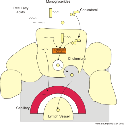
[edit | edit source]The micelles enable the fatty acids and cholesterol to cross the unstirred layer and come in contact with the brush border, where they easily cross the fat-soluble cell membrane. A few smaller free fatty acids transfuse across the cell and out at the baso-lateral border, passing into the capillaries. However most fatty acids enter the smooth endoplasmic reticulum where whey are repackaged into cholemicrons. These are carried out of the cell by exocytosis.
The cholemicrons do not enter the capillaries, but pass instead into the lymphatic system where they are carried to the thoracic duct. The thoracic duct empties into the superior vena cava.
Nucleic Acids
[edit | edit source]RNA and DNA are broken down by pancreatic enzymes and enzymes in the intestinal mucosa.
| Source | Enzyme | Activated by | Substrate | Functions and/or Products |
|---|---|---|---|---|
| Pancreas | Ribonuclease | ... | RNA | Nucleotides |
| Deoxyibonuclease | ... | DNA | Nucleotides | |
| Intestinal mucosa | Nuclease | ... | Nucleic Acids | Purine and pyramidine bases and Pentoses |
The nucleic bases are absorbed by active transport, the pentoses are absorbed with the other sugars.
Vitamins and Minerals
[edit | edit source]Vitamins
[edit | edit source]The fat soluble vitamins A, D, and E are absorbed in the upper small intestine. The factors that cause malabsorption of fat can also affect absorption of these vitamins. Vitamin B12 is absorbed in the ilium and requires to be bound to intrinsic factor, a protein secreted in the stomach, to be absorbed. If intrinsic factor is missing then Vitamin B12 is not absorbed and pernicious anaemia results.
Of the water soluble vitamins, transport of Folate and B12 across the apical membrane are Na+ independent, but the other water soluble vitamins are absorbed by Na+ co-transporters.
Calcium
[edit | edit source]Between 30% and 80% of the body's calcium intake is absorbed. This, and the relationship of Ca++ absorption to the Vitamin D derivative 1,25-dihydroxycholecalciferol is discussed further in the chapter on Ca metabolism. The rate of absorption is dependent on the body's Ca++ needs.
Iron
[edit | edit source]Almost all iron absorption occurs in the ferrous (Fe2) form in the duodenum. The ferric (Fe3) form is converted to ferrous by the enzyme ferric reductase. A protein transporter designated DMT1 is responsible for absorption at the apical surface of the enterocyte. Heme molecules are transported by the HT protein transporter. At the basolateral part of the enterocyte Ferrous ions are transported into the interstital fluid by a transporter called ferroportin (FP).
In the plasma the ferrous form is reconverted back to the ferric form, and bound to an iron transporting protein transferrin (TF).
Water and Electrolytes
[edit | edit source]The small intestine is presented with 9 liters, 2 extrinsic, and 7 intrinsic, of fluid a day for reabsorbtion. In health all but 200cc's are reabsorbed. About 1500 ccs of fluid leave the small intestine and enter the colon.
Large Intestine
[edit | edit source]The junctions between epithelial cells in the large intestine are much tighter than in the small intestine, and this precludes leakage of sodium into the lumen. Most of the fluid and electrolytes are absorbed in the ascending colon.
Although proteins and sugars have usually all been absorbed by the time fluid reaches the large intestine, the colon is capable for absorbing these substrates. Some hard to digest substances, such as beans, can be digested by the colonic bacteria, and these bacteria can even digest small amounts of cellulose.


