Molecular Cloning/Checking of DNA on PAGE


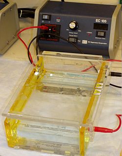
In simple terms Electrophoresis is a procedure which enables the sorting of molecules based on size and charge. Using an electric field, molecules (such as DNA) can be made to move through a gel made of agar. The molecules being sorted are dispensed into a well in the gel material. The gel is placed in an electrophoresis chamber, which is then connected to a power source. When the electric current is applied, the larger molecules move more slowly through the gel while the smaller molecules move faster. The different sized molecules form distinct bands on the gel.[citation needed] The term "gel" in this instance refers to the matrix used to contain, then separate the target molecules. In most cases, the gel is a crosslinked polymer whose composition and porosity is chosen based on the specific weight and composition of the target to be analyzed. When separating proteins or small nucleic acids (DNA, RNA, or oligonucleotides) the gel is usually composed of different concentrations of acrylamide and a cross-linker, producing different sized mesh networks of polyacrylamide. When separating larger nucleic acids (greater than a few hundred bases), the preferred matrix is purified agarose. In both cases, the gel forms a solid, yet porous matrix. Acrylamide, in contrast to polyacrylamide, is a neurotoxin and must be handled using appropriate safety precautions to avoid poisoning. Agarose is composed of long unbranched chains of uncharged carbohydrate without cross links resulting in a gel with large pores allowing for the separation of macromolecules and macromolecular complexes.[citation needed] "Electrophoresis" refers to the electromotive force (EMF) that is used to move the molecules through the gel matrix. By placing the molecules in wells in the gel and applying an electric field, the molecules will move through the matrix at different rates, determined largely by their mass when the charge to mass ratio (Z) of all species is uniform, toward the anode if negatively charged or toward the cathode if positively charged.
In the case of nucleic acids, the direction of migration, from negative to positive electrodes, is due to the naturally-occurring negative charge carried by their sugar-phosphate backbone. Double-stranded DNA fragments naturally behave as long rods, so their migration through the gel is relative to their size or, for cyclic fragments, their radius of gyration. Single-stranded DNA or RNA tend to fold up into molecules with complex shapes and migrate through the gel in a complicated manner based on their tertiary structure. Therefore, agents that disrupt the hydrogen bonds, such as sodium hydroxide or formamide, are used to denature the nucleic acids and cause them to behave as long rods again. Gel electrophoresis of large DNA or RNA is usually done by agarose gel electrophoresis. See the "Chain termination method" page for an example of a polyacrylamide DNA sequencing gel. Characterization through ligand interaction of nucleic acids or fragments may be performed by mobility shift affinity electrophoresis. Electrophoresis of RNA samples can be used to check for genomic DNA contamination and also for RNA degradation. RNA from eukaryotic organisms shows distinct bands of 28s and 18s rRNA, the 28s band being approximately twice as intense as the 18s band. Degraded RNA has less sharpely defined bands, has a smeared appearance, and intensity ratio is less than 2:1.
The DNA to be separated can be prepared in several ways before separation by electrophoresis. In the case of large DNA molecules, the DNA is frequently cut into smaller fragments using a DNA restriction endonuclease (or called restriction enzyme). In other instances, such as PCR amplified samples, enzymes present in the sample that might affect the separation of the molecules are removed through various means before analysis. Once the DNA is properly prepared, the samples of the DNA solution are placed in the wells of the gel and a voltage is applied across the gel for a specified amount of time.
The DNA fragments of different lengths are visualized using a fluorescent dye specific for DNA, such as ethidium bromide. The gel shows bands corresponding to different DNA molecules populations with different molecular weight. Fragment size is usually reported in "nucleotides", "base pairs" or "kb" (for thousands of base pairs) depending upon whether single- or double-stranded DNA has been separated. Fragment size determination is typically done by comparison to commercially available DNA markers containing linear DNA fragments of known length.
The types of gel most commonly used for DNA electrophoresis are agarose (for relatively long DNA molecules) and polyacrylamide (for high resolution of short DNA molecules, for example in DNA sequencing). Gels have conventionally been run in a "slab" format such as that shown in the figure, but capillary electrophoresis has become important for applications such as high-throughput DNA sequencing. Electrophoresis techniques used in the assessment of DNA damage include alkaline gel electrophoresis and pulsed field gel electrophoresis. The measurement and analysis are mostly done with a specialized gel analysis software. Capillary electrophoresis results are typically displayed in a trace view called an electropherogram.
Visualization of DNA by ethidium bromide (EtBr) and otherdyes
[edit | edit source]The most common dye used to make DNA or RNA bands visible for agarose gel electrophoresis is ethidium bromide, usually abbreviated as EtBr. It fluoresces under UV light when intercalated into DNA (or RNA). By running DNA through an EtBr-treated gel and visualizing it with UV light, any band containing more than ~20 ng DNA becomes distinctly visible. EtBr is a known mutagen, and safer alternatives are available. Even short exposure of nucleic acids to UV light causes significant damage to the sample. UV damage to the sample will reduce the efficiency of subsequent manipulation of the sample, such as ligation and cloning. If the DNA is to be used after separation on the agarose gel, it is best to avoid exposure to UV light by using a blue light excitation source such as the Xcitablue UV to blue light conversion screen from Bio-Rad or Dark Reader from Clare Chemicals. A blue excitable stain is required, such as one of the SYBR Green or GelGreen stains. Blue light is also better for visualization since it is safer than UV (eye-protection is not such a critical requirement) and passes through transparent plastic and glass. This means that the staining will be brighter even if the excitation light goes through glass or plastic gel platforms. SYBR Green I is another dsDNA stain, produced by Invitrogen. It is more expensive, but 25 times more sensitive, and possibly safer than EtBr, though there is no data addressing its mutagenicity or toxicity in humans. SYBR Safe is a variant of SYBR Green that has been shown to have low enough levels of mutagenicity and toxicity to be deemed nonhazardous waste under U.S. Federal regulations. It has similar sensitivity levels to EtBr, but, like SYBR Green, is significantly more expensive. In countries where safe disposal of hazardous waste is mandatory, the costs of EtBr disposal can easily outstrip the initial price difference, however. Since EtBr stained DNA is not visible in natural light, scientists mix DNA with negatively charged loading buffers before adding the mixture to the gel. Loading buffers are useful because they are visible in natural light (as opposed to UV light for EtBr stained DNA), and they co-sediment with DNA (meaning they move at the same speed as DNA of a certain length). Xylene cyanol and Bromophenol blue are common dyes found in loading buffers; they run about the same speed as DNA fragments that are 5000 bp and 300 bp in length respectively, but the precise position varies with percentage of the gel. Other less frequently used progress markers are Cresol Red and Orange G which run at about 125 bp and 50 bp, respectively. Visualization can also be achieved by transferring DNA to a nitrocellulose membrane followed by exposure to a hybridization probe. This process is termed Southern blotting.
Running buffer
[edit | edit source]There are a number of buffers used for agarose electrophoresis. The most common being: Tris/Acetate/EDTA (TAE), Tris/Borate/EDTA (TBE) and lithium borate (LB). TAE has the lowest buffering capacity but provides the best resolution for larger DNA. This means a lower voltage and more time, but a better product. LB is relatively new and is ineffective in resolving fragments larger than 5 kbp; However, with its low conductivity, a much higher voltage could be used (up to 35 V/cm), which means a shorter analysis time for routine electrophoresis. As low as one base pair size difference could be resolved in 3 % agarose gel with an extremely low conductivity medium (1 mM Lithium borate).
How to analyze the gel
[edit | edit source]After electrophoresis the gel is illuminated with an ultraviolet lamp (usually by placing it on a light box, while using protective gear to limit exposure to ultraviolet radiation) to view the DNA bands. The ethidium bromide fluoresces reddish-orange in the presence of DNA. The DNA band can also be cut out of the gel, and can then be dissolved to retrieve the purified DNA. The gel can then be photographed usually with a digital or polaroid camera. Although the stained nucleic acid fluoresces reddish-orange, images are usually shown in black and white (see figures).
Gel electrophoresis research often takes advantage of software-based image analysis tools, such as ImageJ.
| 1 | 2 | 3 |
|---|---|---|
 |
 |
 |
