Planet Earth/7d. The Origin of Sex
Asexual Reproduction
[edit | edit source]


Bacteria belong to a group of unicellular organisms, called the prokaryotes that exhibit a cellular membrane and wall that surrounds a free-floating circular DNA molecule held in a cytoplasm fluid with various proteins produced by ribosome RNA, and coded by the DNA molecule. The cellular membrane and wall are constructed by lipids and proteins, produced by RNA from amino acids and carbohydrates that most bacteria will consume from the outside environment. Some bacteria exhibit long, whip-like protrusions from their cellular wall that aids cellular locomotion, called flagella, while others are able to secrete calcium carbonate forming a hard-skeletal cellular wall. Prokaryotes mostly reproduced by asexual reproduction by binary fission, which is where the DNA molecule replicates a nearly identical functional copy and the cellular membrane pitches off into two cells (cytokinesis), each containing a DNA molecule. Most prokaryotes replicate by this simple process of asexual reproduction. Hence, a single cell can reproduce very quickly by doubling. As long as there is sufficient amino acids and carbohydrates in the environment and or proteins, bacteria can quickly reproduce to large numbers from a single cell. This doubling allows bacterial colonies to grow rapidly in population size.
Individual bacterial cells do eventually die, which is highly dependent on environment conditions. For example, if the environment becomes too harsh, such as in lacking nutrients, high-water temperatures, acidic or basic water pH, high salinity, short wave length electromagnetic radiation, nuclear radiation or high oxidation, the cells will perish. This means that early bacterial populations were held back by the conditions of the external environment and availability of nutrients vital for life, which early on were produced naturally from the Earth’s atmosphere and oceans, but in limited supply. If a large bacterial population faced a harsh environment, many of these single celled organisms would perish and die, leaving fresh sources of nutrients behind of complex organic molecules by their decaying cellular masses. If just a single living cell was able to survive this harsh environment, there was a great benefit to it, as there would be amble supply of nutrients for the survivors. The trait that makes a living organism better at surviving in an environment is called an adaptation. These adaptations likely originated by the randomization that is brought about during the replication of DNA in the cell.

DNA is short for deoxyribonucleic acid a large organic molecule composed of two polynucleotide chains that coils around each other forming a double helix. Each molecule of DNA is unique as to each molecule, and carries very large strains of individual unique nucleotides, which are composed of one of four nucleobases (cytosine [C], guanine [G], adenine [A], and thymine [T]), each connected to deoxyribose and a phosphate group, that provides the sugar-phosphate backbone to DNA. The double helix strains are held together by weak hydrogen bonds between the nucleobases, with adenine [A] pairing with thymine [T], and cytosine [C] pairing with guanine [G]. These pairings are easily broken during the first step of replication of the DNA molecule, where the double helix is “unzipped,” using an enzyme called helicase. The two single loose strands of DNA will act as templates to make new copies.

One strand is called the leading strand, which has a 3′ prime end capped with a terminal hydroxyl group, while the lagging strand has a 5’ prime end capped with a terminal phosphate group. Both will act as templates to build a copy of each side or strand of the original DNA. The leading strand will build onto this template using a primer RNA molecule that binds to the leading strand at the 3′ prime end and moves toward the 5’ prime end. DNA polymerase binds to the leading strand and moves down the template adding complementary nucleotide bases (A, C, G, and T) as it goes along the strand building a nearly identical copy of the original lagging strand of DNA. This is called continuous replication. It was often thought that the original lagging strand of DNA was built along the same fashion. We today know, that it does something different thanks to a brilliant woman from Japan.

Tsuneko Okazaki (岡崎 恒子) was born in Japan in 1933, and witness the horror of the Second World War while in grade school, the lasting effects of which would haunt the rest of her life. In the aftermath of the devastating war, she enrolled in post-secondary school to study biology earning a PhD in Japan, during a time in which woman were first permitted to earn advanced degrees at a university. She loved working in the lab, studying the early cell division in frog and sea urchin eggs, and understanding how these early cells replicate and grow. She fell in love with Reiji Okazaki (岡崎 令治), a fellow scientist who also studied in the lab, Reiji Okazaki had experienced the death and destruction of war as well, having been exposed to the radiation fallout of the Hiroshima nuclear bombing as a child. The two married, moved to the United States to continue their research, and begin to focus on the common bacteria Escherichia coli, that lives in the intestine of many animals and humans. Their work demonstrated something unusual in how the lagging strand of DNA is copied. The lagging strand of DNA is built piecemeal from fragments that are added to the opposite side following the same direction as the leading strand, with the first parts built near the unzipping, and working down toward the 5’ prime end. These fragments (which are called Okazaki fragments) are added using RNA primers, which will need to be replaced. Once these fragments are placed next to their corresponding bases (A with T, C with G), an enzyme called exonuclease pulls out the temporary primers and fills the gaps with any missing nucleotides. This process is called discontinuous replication.
Once each side of the original DNA molecules has resulted in a new paired DNA molecule, the enzyme DNA ligase binds up the sequence and results in two new DNA molecules, each with one half of the original DNA molecule and one half with a copied DNA molecule. The process is complicated, and errors can be introduced during this delicate process of cellular replication. If exposed to nuclear radiation these processes of replication can stop, or mismatches can occur caused by the breakage of these delicate linkages inside each cell. In a bacterial colony, or in the living tissue of a human being, such mistakes can lead to abnormal cells. Most of these abnormal cells will fail, but some can become cancerous, copying abnormally until they replace healthy cells.
The high exposure of nuclear radiation of the Hiroshima bomb dramatical increased this risk to Reiji Okazaki, while the couple moved back to Japan, celebrated the birth of their two children, and success of their international research, Reiji was becoming ill. In 1975 he died of cancer, at the age of only 44, leaving behind his beloved wife and two small children. Tsuneko continued her research, and advocated for better resources for woman scientists in Japan, as she was suddenly thrust into the difficult life of raising two children alone and carrying out her intense scientific experiments. Nevertheless, she persevered, by making new discoveries in the field of genetics and how cells divide, and leading further research into more complex organisms.
The complexity of the replication of DNA always leads to the possibility that mistakes or differences will appear in the strands of the four possible nucleobases (cytosine [C], guanine [G], adenine [A], or thymine [T]). For example, where a cytosine might accidentally be replaced with a guanine, these mistakes introduce variation, which are called mutations. Mutations can be bad or in more rare cases they can be advantageous, allowing a cell to survive in a slightly harsher environment for example. In the early Earth, with only bacterial single cells replicating in such a way, any innovation only came from the random likelihood of a mistake turning out good. This process resulted in the continued trial and error method for any change or advancement within individual cells. Most cells would be near perfect copies or clones of the original cell. If the environment changed harshly, and the bacteria did not escape, the entire colony of cells would nearly all perish. Those that survived death could go on replicating.
The Advent of Sex
[edit | edit source]One of the greatest innovations in the evolution of life on planet Earth was the origin of sexual reproduction. Most bacteria reproduce by making copies, or clones of themselves that closely resemble the original cell, asexual reproduction. This allowed bacterial populations to grow in great numbers, but also die in great numbers when the conditions for life change. This is because each descendant cell is nearly identical to the original cell, and there is little genetic variation within the population.


One of the most amazing abilities that bacteria developed was the ability to share genetic information between cells, either as fragments of RNA or DNA that could be used to communicate between individual cells. This process of reproduction is an early form of sexual reproduction. Modern bacteria do this through the use of a pilus (also called a sex pilus), which is a long tube-like appendage that can carry fragments of novel DNA or RNA and share this with surrounding cells. The pilus can also serve as an anchor point to bind bacteria to other cells, holding them in place, and likely used to provide genetic communication between cells. This additional DNA or RNA molecules can increase genetic diversity within a population of cells more quickly than simple random mutations.
One theory for the origin of viruses, suggests that these fragments of DNA or RNA which encode or alter the bacteria’s original DNA, mutated into forms that code for the replication of more viruses, instead. Indeed, this is very likely the origin of many viruses that exist today, and likely most viruses are miss-coded RNA or DNA strands that continue to infect cells, and altering them in the process. It is remarkable that the vast majority of differences between DNA molecules in a population of individual cells is likely do to the interaction of virial RNA or DNA infections that can change and alter the genetic code of these cells. Much of this information maybe excessive and non-informative for the production of proteins or the expression of traits within the cell, but it does enhance the probability of new and novel features that might appear with beneficial results.
If two colonies of bacteria are replicating, the colony that shares genetic information between cells will have a major advantage compared to the one that has to rely on the randomness of mutations and mistakes during replication. Such early networks of communication between cells became vitally important during the early stages of life on Earth, as cells begin to work as a network of individual cells, rather than individualistically. This type of sexual reproduction is fundamentally very different than that observed by more complex single celled organisms, as well as plants and animals, but both serve the purpose of increasing genetic variability within a population. The ability to share genetic information significantly improves the outcome of adaptive individuals that are able to survive changes to the environmental conditions.
The Origin of Complexity of Single Cells
[edit | edit source]
In the laboratories at the University of Chicago, Stanley Miller’s experiments sparked in the corner of Harold Urey’s lab, a demonstration that the vital ingredients required for primitive lifeforms could be easily produced in the ancient atmosphere and ocean of Earth. These amino acids and carbohydrates would have allowed primitive early bacteria to thrive in this primordial world. However, with rising populations inevitably these rapidly reproducing bacteria would use up all of the limited input of these natural resources. As such, life was precious as it depended on the amount of nutrients provided by the natural processes, coupled with the harsh environment of an early Earth. It was no surprise that some cells during this time turned to feed on other cells. This was the origin of heterotrophic organisms, which take in organic substances from other living cells. In addition to the nutrients from natural occurring chemical reactions and other living or decaying organisms, many of these early microscopic single-celled lifeforms utilized the primordial atmospheric gasses for a type of respiration, or energy source, principally CO2, SO2 and NO2. These early cellular lifeforms are called the Archaea, or archaebacteria, from the Greek arkhaios meaning primitive. They thrive today in environments that lack free oxygen, and many are also often identified as extremophiles, which are bacteria that can exist in harsh environmental conditions, such as anoxic or euxinic waters, deep underground, hot sulfur springs, or extreme warm or cold climates that may have been more common during the Archean period. The three major types of archaebacteria lifeforms can be divided based on how they rely on chemosynthesis, the synthesis of organic compounds by living organisms using energy derived from reactions involving inorganic chemicals only, typically in the absence of sunlight.
First are the methanogenesis-based lifeforms that take advantage of carbon dioxide (CO2), by using it to produced methane CH4 and CO2, through a complex series of chemical reactions in the absence of oxygen. Methanogenesis requires some source of carbohydrates (larger organic molecules containing carbon, oxygen and hydrogen) as well as hydrogen, but these organisms produce methane (CH4) particularly in sediments on sea floor in the dark and deep regions of the oceans. Today they are also found in the guts of many animals. Second are the sulfate-reducing lifeforms that take advantage of sulfur in the form of sulfur dioxide (SO2), by using it to produce hydrogen sulfide (H2S). Sulfate-reducing life forms require a source of carbon, often in the form of methane (CH4) or other organic molecules produced naturally, such as amino acids, as well as sources of sulfur, typically near volcanic vents deep underwater. And finally, the nitrogen reducing lifeforms, which take advantage of nitrogen in the form of nitrogen dioxide (NO2) by using it to produce ammonia (NH4). Nitrogen-reducing life forms also require a source of carbon, often in the form of methane (CH4) or other organic molecules produced naturally (many of these bacterial organisms form a symbiotic relationship with plants). All three types of life-forms exhibit anaerobic respiration, or respiration that does not involve free atoms of oxygen. They all benefit from the input of naturally occurring amino acids, carbohydrates and other complex carbon-based molecules that the Urey-Miller experiment at the University of Chicago demonstrated could form naturally on a primordial Earth, but also taken in organic molecules from other single celled organisms.
Endosymbiosis
[edit | edit source]
Two inquisitive love-struck teenagers were hanging out at the lab pondering the ramifications of the Urey-Miller experiment. Their names were Carl Sagan and Lynn Margulis. Sagan had blazed through high school graduating at the young age of 16, and had enrolled at the University of Chicago, where as an undergraduate he was working in the lab of Harold Urey as a young student. Lynn Margulis was a 15-year-old local high school student, interested in science, enrolled in the University of Chicago Laboratory School, when the two met in Chicago. At the end of their undergraduate college experience and dating, the two married in 1957, and had two sons (Dorion Sagan and Jeremy Sagan), while Carl dreamed of extraterrestrial life, switching his degree to astronomy and physics, and later popularized science with his television show. Lynn instead focused on how life became complicated and went on to study biology in graduate school at the University of Wisconsin.

The couple struggled with the demands of academia and raising two boys while trying to conduct research and teaching. At the University of California Lynn Margulis focused on a deceptively simple organism for her doctoral research called Euglena, which is a Eukaryote. Eukaryotes are more complex organisms that have a cell or cells composed of a nucleus enclosed within a membrane that holds the genetic information of DNA (often as chromosomes), as well as a number of organelles within the cell that provide special functions to the cell. Euglena is unique in that it is a mobile type of algae that lives in pond water, swimming around with a flagellum, but also is able to photosynthesize like plants. Most species of Euglena have many photosynthesizing green chloroplasts within cell, which allows them to generate food and energy from carbon dioxide and sunlight. These single celled organisms are an example of an autotroph (also called a primary producer), a life form that can generate its own food from the surrounding inorganic environment.
Chloroplasts, and the advent of carbon dioxide breathers
[edit | edit source]Lynn Margulis realized that the advent of such organisms must have been a major breakthrough for early organisms in Earth’s history. The origin of chloroplasts, and ability for cells to photosynthesize must have completely altered the world. A major scientific discovery she personally made came about in wondering where these chloroplasts within the cells of Euglena came from. What allowed these creatures to be able to photosynthesize in the first place?
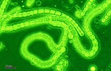
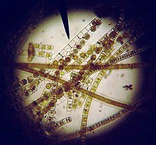
The origin of an organism that is able to take carbon dioxide gas and photons (in addition to traces of ammonia, phosphate, potassium and other nutrients) and build them into the needed raw ingredients (mostly carbohydrates) for food within the cell was a mystery. She searched around for other types of single celled algae that use photosynthesis, and she noticed that some forms of algae are in fact tiny prokaryotic organisms, classified in a group called the blue-green algae, or more appropriately named cyanobacteria. These are bacteria that are able to photosynthesis, often replicating in long strains of tiny cells, that live in the photic zone of the world’s oceans (but can survive nearly everywhere on Earth where there is sunlight).
The occurrence of cyanobacteria prokaryotic organisms with the ability to photosynthesize would completely alter Earth in extraordinary and profound ways. The early Earth when the Archean bacteria dwelled was significantly limited by the environmental conditions and availability of nutrients. The advent of photosynthesis unlocked a vast storage of carbon dioxide directly from Earth’s early atmosphere. Like Mars and Venus, the early atmosphere of Earth was composed of nearly 95% carbon dioxide, this carbon dioxide was the source of carbon in the natural development of organic molecules necessary for life. What this group of single celled prokaryotes acquired was the ability to directly take in carbon dioxide and produce carbohydrates necessary for growth. As long as there was carbon dioxide in the atmosphere the cyanobacteria could flourish and reproduce in vast numbers. Very quickly cyanobacteria became the dominate lifeform on Earth, the fossils of which are preserved because of a unique interaction this process has on the formation of calcium carbonate.

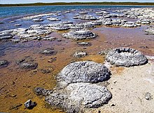
Photosynthesis takes out the carbon from acidic waters in which carbon dioxide gas is dissolved in the ocean water as carbonic acid, and returns oxygen gas to the water. This raises the pH of the water (making it more basic) surrounding the cell, which results in the precipitation of calcium carbonate in marine and freshwater systems. These bacteria increase pH as a result of this photosynthetic activity and also produce extracellular sticky polysaccharide sugars, which act as binding sites for calcium and carbonate ions coating the cell in a protective skeleton. These cells when they die get buried in the subsurface and form calcium carbonate limestones. Ancient limestones and fossil algal mats called stromatolites preserve a record of the first appearance of these organisms during the Archean, about 3 billion years ago, and their steady increase over a great span of time. These cyanobacteria released oxygen, while drawing down carbon dioxide to lower and lower levels in the atmosphere and oceans.
Heterotrophic organisms benefited from these new organisms, as they had a source of nutrients in these rapidly growing populations of cyanobacteria, and they likely consumed them by evaginating their cells around these organisms, releasing their nutrients. The thick carbonate skeletons were an adaptation to protect these organisms from being consumed. As Lynn Margulis watched her study organism, Euglena, dance around under her microscope, with a cell filled with green chloroplast organelles, she wondered if these green chloroplast organelles were just cyanobacteria that instead of being consumed by the cell, were in fact just incorporated within the cellular structure. The advantage was clear, as keeping cyanobacteria alive would provide the whole cell a continued source of carbohydrates. Was this a form of symbiosis, in which each tiny squirming cell of Euglena in actuality a multicellular organism of various species of prokaryotic cells living together? Was eukaryote cells actually made up of multiple species of prokaryotic cells with different functions? This idea was a radically different view of biology, which is called endosymbiosis. She drafted a famed paper proposing the idea entitled “Evolutionary Criteria in Thallophytes: A Radical Alternative” Thallophytes is an abandoned term for primitive fungus and algae, and primitive plants, of which Euglena was a member of. The paper was rejected numerous times, as reviewers argued that she did not have the evidence to back such a radical idea, despite this she was able to persevere and got it published in Science gathering some amazing evidence for symbiotic relationships in all eukaryotic cells.

The full force of evidence backing her idea would come swiftly from the study of another organelle in eukaryotic cells, one that is found inside your own cells – mitochondria. Mitochondria are oval to rod shaped organelles found in most eukaryotic cells. They have a double membrane with an inner layer that forms pockets called cristae. The mitochondria serve an important function in the process of respiration and energy production for more complex eukaryotic cells.
Mitochondria, and the advent of the oxygen breathers
[edit | edit source]During the billions of years of the Archean the amount of available carbon dioxide for photosynthesis in the Earth’s atmosphere was used up by the highly successful cyanobacteria, reducing the Earth’s atmosphere of carbon dioxide being replaced with oxygen. The amount of input of new carbon dioxide was limited to the volcanic activity of the Earth, which was still much more active than today, as the accretionary heat was much greater early in Earth’s history, but still limited. This limited flow of carbon dioxide into the atmosphere meant that it would often get used up by photosynthesizing cyanobacteria in vast algae blooms following volcanic eruptions. The atmosphere was instead filling with a new dangerous gas – oxygen. For most Archaea bacteria, oxygen is a poisonous gas that causes harmful oxidation to the cells and reduces the ability for them to conduct chemosynthesis. What is worst, is that oxygen destroys methane, ammonia, and hydrogen sulfide from the atmosphere and oceans that these prokaryotic cells need to live.

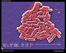
There was one group of prokaryotic organisms that developed the ability to derive energy from the free oxygen now in abundance in the atmosphere and ocean. These were the precursors to the mitochondria found in most eukaryotic cells. Mitochondria converts free oxygen and nutrients into adenosine triphosphate (abbreviated ATP). ATP is how cells store chemical energy that can be used in metabolic activities, such as growth, movement and cellular replication. This process (called aerobic respiration) works by taking free oxygen and using it to convert carbon-based molecules into ATP that can be used for energy later in the cell. This highly efficient method of energy storage allows these types of cells to survive longer in an oxygen-rich environment that was beginning to occur in the early Earth. These organisms, however, still had to find a source of carbon, which they did so by being a heterotrophic organism, that is feeding on other cells. These bacteria are the aerobic prokaryotes. Some examples include the infectious Staphylococcus, a type of bacteria that feeds on animal cells, and causes many bacterial diseases and infections. Staphylococcus is a facultative anaerobe which means that cells can make ATP in the presence of oxygen, but can still survive anoxic environments by switching to anaerobic respiration, which is less efficient. The common gut bacteria Escherichia coli is another example of a prokaryote that exhibits both aerobic and anaerobic respiration. These bacteria survived by taking in oxygen, and respiring carbon dioxide back into the environment.

When Lynn Margulis looked through her microscope watching the tiny moving individual celled Euglena, she could see that they in addition to having chloroplasts, also had mitochondria. These little eukaryotic organisms were a community, each and every one of them like tiny ships carrying prokaryote passengers, some that could photosynthesize and make carbohydrates, while others produced ATP for energy. Inside the cells, these chloroplasts and mitochondria could reproduce asexually independent as these organelles where like a well crewed ship, each type of organelle with a unique beneficial purpose for the cell as a whole. The heart of the ship, was the nucleus, protected by a membrane with its own series of DNA, often paired into chromosomes as twisted treads of double helix strands of DNA. The nucleus was like the captain of these tiny ships. If true, that the individual organelles were individual cooperating prokaryotic crew members, they should have their own genetic information within each organelle. In the 1960s, Margit Nass-Edelson and her husband Sylvian looked at mitochondria organelles under a scanning electron microscope, and discovered they do indeed have DNA within them. Lynn Margulis also documented DNA within the chloroplast organelles. Eukaryotic organisms were a symbiotic group of organisms working together to provide energy and nutrients to the larger cell. The endosymbiosis theory revolutionized biology, demonstrating an important driver in the strength of a community of organisms even at the smallest scales of life, such as a single eukaryotic cell.
Your body is a multiple of millions of individual ships filled with tiny prokaryotic evolved organelles all working together to give you the consciousness to read these pages, to understand these thoughts. Every human is a planet of their own multitudes. Illness and sickness and the consistent battles that rage inside your cells play out, as do the armies of your multitudes, without your own acknowledgment. Keeping you breathing oxygen, transporting your blood, making you crave food and drink, and serving you and your survival. It is odd to think about, that we are not a single one, but multitudes and multitudes of individual cells.
| Previous | Current | Next |
|---|---|---|
