Proteomics/Protein Separations- Electrophoresis/Techniques
Gel Electrophoresis
[edit | edit source]
Gel electrophoresis is a group of techniques used by scientists to separate molecules based on physical characteristics such as size, shape, or isoelectric point. Gel electrophoresis is usually performed for analytical purposes, but may be used as a preparative technique to partially purify molecules prior to use of other methods such as mass spectrometry, PCR, cloning, DNA sequencing, or immuno-blotting for further characterization. (Wikipedia)
Separation
[edit | edit source]The Gel is created by the use of a matrix molecules stacked together to provide separation of molecules. Gel are normally crosslinked polymers normally made of either agarose gel or polyacrylamide. Dependent on the substance being run the composition, pourous nature, and percentage of gel will be varied to provide proper separation. This separation using acrylamide is normally done on proteins, DNA, RNA, oligonucleotides and other smaller molecules due to its less pourous nature. Using different concentration and compositions of aryclamide provide a thicker or thinner matrix which will separate based on size and charge. Separating larger nucleic acids (Large # of nucleic bases) is normally done with agarose gels, gels made from seaweed extract. Again cases of concentration and composition can vary the results of the distance a sunstance travels on a gel. Techniques such as use of stacking gels are employed where the top half of a gel may be more concentrated than a lower portion to provide further separations.
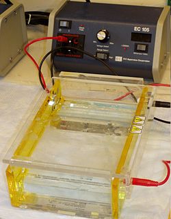
Electrophoresis refers to the electromotive force (EMF) that is used to push or pull the molecules through the gel matrix; by placing the molecules in wells in the gel and applying an electric current, the molecules will move through the matrix at different rates, towards the anode if negatively charged or towards the cathode if positively charged. (Wikipedia) The voltage as well as the time depend on the size of the gel and the type of sample. Caution must be taken to not run the samples too long or the samples may run off the gel. Caution must also be taken with the voltage, voltage too high may burn the gel or damage the samples.
Staining
[edit | edit source]When the gel electrophoresis has completed its run the gel is stained to allow for the bands of sample to be seen and analyzed. Stains that are normally used are Ethidium Bromide, silver, or coomassie blue dye depending on the sample. The gels are cafefully removed and placed directly into staining solution where it will sit overnight to allow for proper staining. Once fully stained bands should appear if samples were run correctly along with a molecular mass marker which should be placed on both ends of the gel to provide proper analysis. Visualization can be done by the naked eye as well as under a UV light to provide easier visualization.
Analysis
[edit | edit source]Bands are then compared to those of the known molecular mass marker on the sides of the gel to determine the approximate mass of the substance as well as separate out bands. A sample that has several bands coming from a single sample can indicate an impure sample. Further analysis can be done on proteins by removing the separted sample from the gel by cutting the band out and using In-gel digestion to remove the protein from the gel for further analysis.
Nucleic acids
[edit | edit source]In the case of nucleic acids, the direction of migration, from negative to positive electrodes, is due to the natural negative charge due to the sugar phosphate backbone of a DNA molecule. Double-stranded DNA fragments are normally long stranded molecules, therefore their migration through the gel is relative to their size of DNA molecules and amount of bases. Single-stranded DNA or RNA tend to fold into tertiary structure due to their wearer single backbone and travel through the gel in a complicated manner. DNA can be denatured by breaking hydrogen bonds and renatured to provide easier and more accurate results frm the gel.
Proteins
[edit | edit source]Proteins vary on many different levels in comparison to DNA. Protein can have different charges and complex shapes, primary, secondary, tertiary, and quaternary structure that make migration through the gel have extremelyt different rates when placing a negative to positive EMF on the sample. Due to these complex structures proteins are usually denatured, or broken down to simple primary structures in the presence of a detergent such as sodium dodecyl sulfate/sodium dodecyl phosphate SDS/SDP which coats the proteins with a negative charge. The amount of SDS bound and used is relative to the size of the protein, this allows the protein to have an overall negative charge and allow for proper migration.
