Radiation Oncology/Thorax/Anatomy
Appearance
|
Front Page: Radiation Oncology | RTOG Trials |
|
|
NSCLC: Main Page | Overview | Anatomy | Screening | Early Stage Operable | Early Stage Inoperable | Locally Advanced Unresectable | Locally Advanced Resectable | Palliation | Brachytherapy | PCI | Miscellaneous | Large cell neuroendocrine | Level I Evidence | |
Lung Anatomy
- See also: general anatomy page at Radiation_Oncology/Anatomy#Thorax
Lung Anatomy
[edit | edit source]
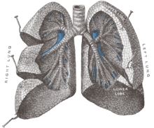
- Lungs: Right and Left
- Lobes:
- Right Lung: Right Upper Lobe (RUL), Right Middle Lobe (RML), Right Lower Lobe (RLL). Separated by oblique fissure and horizontal fissure
- Left Lung: Left Upper Lobe (LUL), Left Lower Lobe (LLL). Separated by oblique fissure. Lingula corresponds anatomically to RML, but is part of the LUL
- Segments
| Right | Segments | Left | Segments |
|---|---|---|---|
| RUL | 3 | LUL | 5 |
| RML | 2 | ||
| RLL | 5 | LLL | 5 |
- Bronchi
- Trachea divides into right main bronchus and left main bronchus, supplying right and left lung respectively
- Each main bronchus divides into secondary (lobar) bronchi, depending on how many lobes each lung has (3 lobar bronchi on the right, 2 lobar bronchi on the left)
- Each secondary bronchus divides into tertiary (segmental) bronchi, depending on the number of segments each lobe has
- Tertiary bronchi divide into bronchioles
- Lung volumes are typically evaluated by spirometry
- Total Lung Capacity (TLC): Volume of lung at the end of maximal inspiration. Depends on age, height, weight, sex, altitude, etc. Normally 4-6 L; women have 20-25% lower TLC than men
- Forced Vital Capacity (FVC): Volume of air that can be forced out after maximal inspiration
- Forced Expiratory Volume in 1 second (FEV1): Volume of air that can be forced out in one second
- FEV1/FVC (FEV1%): Ratio of FEV1 to FVC, i.e. the proportion of Vital Capacity that can be expelled during one second. Typically 75-80%
Lymph node stations
[edit | edit source]- Conceptual organization
- Intrapulmonary (along secondary bronchi)
- Hilar (bronchopulmonary, along main bronchi)
- Mediastinal
- Supraclavicular
IASLC Classification; 2009
[edit | edit source]- 2009 Full text PMID 19357537 -- "The IASLC lung cancer staging project: a proposal for a new international lymph node map in the forthcoming seventh edition of the TNM classification for lung cancer." (Rusch VW, J Thorac Oncol. 2009 May;4(5):568-77.)
- Proposed lymph node map which reconciles Japanese, European, and North American maps for AJCC/UICC 7th edition
- N2 nodes: (all lie within the mediastinal pleural envelope)
- 1 - low cervical, supraclavicular, and sternal notch nodes
- Upper border: lower margin of cricoid cartilage
- Lower border: clavicles bilaterally and in the midline the upper border of manubrium
- 2 - upper paratracheal nodes
- Upper border: apex of lung and pleural spaces and in the midline the upper border of manubrium
- Lower border 2R: intersection of caudal margin of innominate vein with the trachea
- Lower border 2L: superior border of the aortic arch
- 3A - prevascular nodes
- Upper border: apex of chest
- Lower border: carina
- Anterior border: sternum
- Posterior border: anterior border of SVC
- 3P - retrotracheal nodes
- Upper border: apex of chest
- Lower border: carina
- 4 - lower paratracheal nodes
- Upper border 4R: intersection of caudal margin of innominate vein with the trachea
- Upper border 4L: superior border of the aortic arch
- Lower border 4R: lower border of azygos vein
- Lower border 4L: upper rim of the left main pulmonary artery
- 5 - subaortic (AP window) - lateral to the ligamentum arteriosum
- Upper border: lower border of aortic arch
- Lower border: upper rim of left main pulmonary artery
- 6 - para-aortic nodes - lie anterior and lateral to the ascending aorta and aortic arch
- Upper border: upper border of aortic arch
- Lower border: lower border of aortic arch
- 7 - subcarinal - caudal to the carina but not associated with the lower lobe bronchi
- Upper border: carina
- Lower border: upper border of lower lobe bronchus
- 8 - paraesophageal (below carina) - adjacent to the wall of the esophagus, excluding subcarinal nodes
- Upper border: upper border of lower lobe bronchus
- Lower border: diaphragm
- 9 - pulmonary ligament - lie within the pulmonary ligament
- Upper border: inferior pulmonary vein
- Lower border: diaphragm
- 1 - low cervical, supraclavicular, and sternal notch nodes
- N1 nodes: (intrapulmonary and hilar)
- 10 - hilar nodes - immediately adjacent to mainstem bronchus and hilar vessels
- 11 - interlobar nodes
- 12 - lobar
- 13 - segmental
- 14 - subsegmental
AJCC/UIC Classification; 1997
[edit | edit source]- 1997 PMID 9187199 Full text — "Regional lymph node classification for lung cancer staging." Mountain CF et al. Chest. 1997 Jun;111(6):1718-23.
- N2 nodes: (all lie within the mediastinal pleural envelope)
- 1 - highest mediastinal nodes - nodes lying above a horizontal line at the upper rim of the brachiocephalic (left innominate) vein where it ascends to the left, crossing in front of the midline of the trachea. Superior border at sternal notch.
- 2 - upper paratracheal nodes - nodes lying above a horizontal line drawn tangential to the upper margin of the aortic arch and below the inferior boundary of #1 nodes
- 3A and 3P - prevascular (3A) and retrotracheal (3P) nodes -
- 4 - lower paratracheal nodes - lie lateral to the midline of the trachea between a horizontal line drawn tangential to the upper margin of the aortic arch and a line extending across the R (or L) main bronchus
- 5 - subaortic (AP window) - lateral to the ligamentum arteriosum or the aorta or L pulmonary artery and proximal to the first branch of the L pulmonary A
- 6 - para-aortic nodes - lie anterior and lateral to the ascending aorta and aortic arch beneath a line tangential to the upper margin of the aortic arch
- 7 - subcarinal - caudal to the carina but not associated with the lower lobe bronchi or arteries within the lung
- 8 - paraesophageal (below carina) - adjacent to the wall of the esophagus, excluding subcarinal nodes
- 9 - pulmonary ligament - lie within the pulmonary ligament
- N1 nodes: (intrapulmonary and hilar)
- 10 - hilar nodes
- 11 - interlobar nodes
- 12 - lobar
- 13 - segmental
- 14 - subsegmental
Graphic depiction of nodal stations
[edit | edit source]Using the 2009 IASCL Classification:
- 2013 No PMID (journal not yet indexed) Abstract -- "Computed tomographic atlas for the new international lymph node map for lung cancer: A radiation oncologist perspective" (Lynch R, Pract Radiat Oncol. 2013 Jan;3(1):54-66)
Using prior classification:
- Mass. General, 2007 PMID 10701624 Full text -- "CT depiction of regional nodal stations for lung cancer staging." (Ko JP, AJR Am J Roentgenol. 2000 Mar;174(3):775-82.)
- University of Michigan thoracic lymph node atlas, 2005 - PMID 16111586 -- "CT-based definition of thoracic lymph node stations: an atlas from the University of Michigan." (Chapet O, Int J Radiat Oncol Biol Phys. 2005 Sep 1;63(1):170-8.)
- Netherlands; Chest Lymph Node Map; 2010 Graphical Update from Rijnland Hospital in Leiderdorp, the Netherlands (Radiologyassistant.NL)
