Sensory Systems/Owls
Birds: Anatomy and Physiology of the Owl's Visual and Auditory System
[edit | edit source]Introduction
[edit | edit source]Owls are nocturnal birds belonging to the order of Strigiformes. They possess extraordinary adaptations that enable them to thrive in low-light conditions. Their large tubular eyes, filled with light-sensitive cells, allow them to navigate almost effortlessly in near-total darkness. Furthermore, their remarkable auditory system, which is highly intricate and distinct, enables them to function effectively even in complete darkness. This exceptional auditory system incorporates specialized feathers that form a parabolic facial disc, adjustable operculum or flaps, and, in certain species, asymmetrical ear openings (where one ear is located higher than the other) which are facing forward and slightly to the side. Through this unique system, owls' brains can create an auditory representation of their surroundings, aiding them in locating their prey.[1]

Visual System
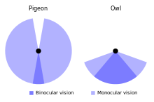
[edit | edit source]Among the various features possessed by owls, their eyes stand out as particularly remarkable. They are large and positioned towards the front, and account for a significant proportion of the owl's body weight, ranging from one to five percent depending on the species. The forward-facing nature of their eyes, which contributes to their "wise" appearance, also grants them a broad range of binocular vision. This means that owls can perceive objects in three dimensions, encompassing height, width, and depth, much like humans. Owls have a field of view spanning approximately 110 degrees, with around 70 degrees comprising binocular vision. In contrast, humans have a field of view that encompasses 180 degrees, with approximately 140 degrees involving binocular vision.

To compensate for its limited field of view, the owl possesses the remarkable ability to rotate its head up to 270 degrees both left and right from its forward-facing position. Furthermore, it can even rotate its head almost completely upside down.[2]


The size of owl eyes is optimized to enhance their effectiveness, particularly in situations with limited lighting. Interestingly, the eyes of an owl are not spherical, but elongated tubes. These tubular eyes are supported and fixed in position by bony structures known as sclerotic rings within the skull. Consequently, owls lack the ability to "roll" or rotate their eyes, limiting their gaze to a fixed forward direction.[2]
The owl's eyes possess an exceptionally large cornea, which is the transparent outer layer of the eye, as well as a sizable pupil located at the center. The larger cornea facilitates a larger pupil size, leading to an increased number of photons reaching the retina, which is the light-sensitive tissue responsible for image formation. As a result, visual sensitivity is enhanced.[3]
The iris, a colored membrane situated between the cornea and lens, regulates the size of the pupil. When the pupil expands, it allows a greater amount of light to pass through the lens and reach the extensive retina. Within the retina, light-sensitive cells serve as receptors and contribute to the formation of images[1]. Similar to humans, owls possess two distinct types of light-sensitive cells in their retinas: rods, responsible for detecting light and movement, and cones, which enable color differentiation. However, there is a significant difference in the ratio of rods to cones between owls and humans. While humans typically have approximately 20 rods for every cone, owls exhibit a ratio closer to 30 to one. This discrepancy grants owls exceptional capabilities in detecting movement, even in low-light conditions.
Beyond the rod-rich retina, there exists an additional layer known as the tapetum lucidum behind an owl's eye. This specialized layer captures any light that penetrates through the retina and reflects it back to the highly sensitive rods. When combined, all of these adaptations result in remarkable low-light sensitivity, with some owl eyes being potentially up to 100 times more adept than ours.[4]
Although it is commonly believed that owls are blind in bright light due to their exceptional night vision, this assumption is false. Owls possess pupils with a wide range of adjustment, allowing them to regulate the amount of light that reaches their retinas effectively. In fact, certain owl species can even perceive bright light better than humans.
To safeguard their eyes, owls possess three eyelids. They have the typical upper and lower eyelids, with the upper eyelid closing during blinking and the lower eyelid rising when the owl is asleep. Additionally, owls possess a third eyelid known as a nictitating membrane. This thin layer of tissue diagonally crosses the eye from the inside to the outside, serving to clean and protect the eye's surface.[2]
Due to the significant size of their "tubular" eyes and being firmly secured by a bony sclerotic ring, owls possess very limited mobility of their eyes[5]. However, in order to overcome this constraint and compensate for their relatively narrow field of view, owls have developed the remarkable ability to smoothly and rapidly swivel their heads laterally by 270 degrees and vertically by 90 degrees.
Auditory system
[edit | edit source]
They possess a well-developed auditory system due to their nocturnal nature, with their ears situated on the sides of their head, behind their eyes, and concealed by the feathers of the facial disc. The apparent "Ear Tufts" found in certain species are actually decorative feathers and not functional ears.[1]
An owl's range of audible sounds is comparable to that of humans. However, their hearing becomes remarkably acute, especially at frequencies of 5 kHz and above. This heightened sensitivity allows them to perceive the slightest movements of their prey within leaves or undergrowth, maximizing their hunting accuracy within the frequency range of 4 to 8 kHz.[1][6]

The sound localization in Owl’s involves utilizing two binaural cues: the interaural time difference (ITD) and the interaural intensity difference (IID).

The utilization of continuous time differences imposes limitations on the range of sounds that can be accurately located, and it also places exceptional demands on the auditory systems of owls. One issue is the presence of phase ambiguity, as ongoing time differences are determined by the phase delay between the two ears. When owls rely solely on low frequencies for sound localization, the maximum time delay, occurring when the sound source is directly opposite one ear, always results in a phase delay of less than 180°. Consequently, each phase delay corresponds to a distinct interaural time difference (ITD) and a specific spatial position. However, with higher frequencies, phase delays exceeding 180° can occur, making it impossible to determine from the ongoing waveform whether the sound is leading in the left or right ear, or leading in one ear by more than one period of the sound frequency. Therefore, for high-frequency tones, the ITDs generated at multiple spatial positions may yield the same relative phase at both ears.
Additionally, the process of sound localization is based on ongoing time differences, as auditory neurons are unable to encode the phase of extremely high-frequency sounds.[7]
Certain owl species possess ear openings that are placed asymmetrically, meaning one ear is positioned higher than the other[6]. This asymmetry creates distinct auditory directional sensitivity patterns for high frequencies, resulting in differences in sound elevation perception between the two ears. As a result, owls are able to localize sounds in the vertical plane by comparing the intensity and spectral composition of sound captured by each ear. In simpler terms, when an owl hears a noise, it can determine the source of the sound by analyzing the minuscule time difference in which it reaches the left and right ears. This time difference, known as the interaural time difference, can be as short as 10 millionths of a second.[1][6]
The strategic arrangement of feathers on the barn owl's facial periphery forms a specialized disc that effectively captures and concentrates sound, akin to the external ears of humans. As sound waves traverse the owl's ear canal, they ultimately reach the eardrum, pass through the ossicles, and enter the inner ear, enabling the owl to precisely determine the whereabouts of its prey. Given that owls rely on their remarkable auditory abilities to track and capture prey, comprehending the intricate structures within the barn owl's ear that facilitate sound transmission is of utmost significance.[1][8][9]
The basilar membrane of the barn owl consists of two primary components: the vestibular part and the tympanic part. In the vestibular part, the basilar membrane is composed of supporting cells, along with a limited number of border cells located at the lower edge of the membrane. Towards the outermost section of the papilla, the basilar membrane is comparatively thin, while it gradually thickens into a fibrous mass as it approaches the innermost section.[9]
In the owl's brain, the signals indicating left, right, up, and down are rapidly integrated to form a cohesive mental image of the spatial location of the sound source. Research conducted on owl brains has unveiled a remarkable complexity in the medulla, the region associated with hearing, surpassing that of other avian species. For instance, it is estimated that a Barn Owl's medulla contains a minimum of 95,000 neurons, three times the number found in a Crow.[6]

Auditory processing pathways in the Owl’s brain
[edit | edit source]The auditory nerve fibers provide innervation to two main cochlear nuclei within the brainstem: the cochlear nucleus magnocellularis the cochlear nucleus angularis. The neurons in the nucleus magnocellularis exhibit phase-locking but are relatively insensitive to variations in sound pressure. On the other hand, the neurons in the nucleus angularis show poor or no phase-locking but are sensitive to sound pressure fluctuations. These two nuclei serve as the initial points for separate yet parallel pathways to the inferior colliculus. The pathway from the nucleus magnocellularis processes interaural time differences (ITDs), while the pathway from the nucleus angularis processes interaural intensity differences (IIDs).
The first location of binaural convergence in the temporal pathway is the nucleus laminaris, which is comparable to the mammalian medial superior olive. It uses coincidence detection and neuronal delay lines to detect and encode ITDs. Laminaris neurons fire most intensely when phase-locked impulses from both ears coincide at the neuron. As a result, the nucleus laminaris functions as a delay-line coincidence detector, converting the sound's travel time into a map of interaural time delays. The anterior lateral lemniscal nucleus and the core of the inferior colliculus' central nucleus receive projections from the nucleus laminaris' neurons.
The posterior lateral lemniscal nucleus, which is similar to the lateral superior olive in mammals, functions as the site of binaural convergence and processes IIDs in the sound level pathway. The neurons are inhibited when the contralateral ear is stimulated, whereas they are excited when the ipsilateral ear is stimulated. The difference between the strength of the inhibitory and excitatory inputs determines the level of excitation and inhibition, which in turn affects how quickly lemniscal nucleus neurons fire. Thus, these neurons' response reflects how ‘loudly each ear is hearing the sound’.
At the lateral shell of the inferior colliculus's central nucleus, the temporal and sound pressure pathways come together. The lateral shell then sends a signal to the external nucleus, where each neuron has a specific receptive field that only responds to sounds coming from that area of space. Only binaural signals with ITD and IID properties similar to those produced by a sound source in the neuron's receptive field will elicit a response from these neurons. Therefore, these space-specific neurons' receptive fields result from their tuning to particular ITD and IID combinations within a constrained range. The positions of receptive fields in space are thus projected in an isomorphic manner onto the anatomical locations of the neurons by these space-specific neurons, resulting in a map of auditory space.[10]
References
[edit | edit source]- ↑ a b c d e f Sieradzki, Alan (2023-03-08), Mikkola, Heimo (ed.), "Designed for Darkness: The Unique Physiology and Anatomy of Owls", Owls - Clever Survivors, IntechOpen, doi:10.5772/intechopen.102397, ISBN 978-1-80355-390-0, retrieved 2023-08-05
- ↑ a b c Lewis, Deane. "Owl Eyes & Vision". The Owl Pages. Retrieved 2023-08-05.
- ↑ Lisney, Thomas J.; Iwaniuk, Andrew N.; Bandet, Mischa V.; Wylie, Douglas R. (2012). "Eye Shape and Retinal Topography in Owls (Aves: Strigiformes)". Brain, Behavior and Evolution. 79 (4): 218–236. doi:10.1159/000337760. ISSN 0006-8977.
- ↑ Kathryn (2022-03-04). ""Owl" Be Seeing You: Amazing Facts About Owl Eyes". American Bird Conservancy. Retrieved 2023-08-05.
- ↑ Steinbach, Martin J.; Money, K.E. (1973-04). "Eye movements of the owl". Vision Research. 13 (4): 889–891. doi:10.1016/0042-6989(73)90055-2. ISSN 0042-6989.
{{cite journal}}: Check date values in:|date=(help) - ↑ a b c d Lewis, Deane. "Owl Ears & Hearing". The Owl Pages. Retrieved 2023-08-05.
- ↑ Volman, S. F. (1994), Davies, Mark N. O.; Green, Patrick R. (eds.), "Directional Hearing in Owls: Neurobiology, Behaviour and Evolution", Perception and Motor Control in Birds: An Ecological Approach, Berlin, Heidelberg: Springer, pp. 292–314, doi:10.1007/978-3-642-75869-0_17, ISBN 978-3-642-75869-0, retrieved 2023-08-05
- ↑ Pena, J. L.; DeBello, W. M. (2010-01-01). "Auditory Processing, Plasticity, and Learning in the Barn Owl". ILAR Journal. 51 (4): 338–352. doi:10.1093/ilar.51.4.338. ISSN 1084-2020.
- ↑ a b Smith, Catherine A.; Konishi, Masakazu; Schuff, Nancy (1985-03-01). "Structure of the Barn Owl's (Tyto alba) inner ear". Hearing Research. 17 (3): 237–247. doi:10.1016/0378-5955(85)90068-1. ISSN 0378-5955.
- ↑ Knudsen, Eric I.; Konishi, Masakazu (1978-05-19). "A Neural Map of Auditory Space in the Owl". Science. 200 (4343): 795–797. doi:10.1126/science.644324. ISSN 0036-8075.
